

Preeclampsia is one of the leading causes of maternal and perinatal death worldwide and affects 2-5% of all pregnancies.1-2 It’s a frightening condition because it can come on suddenly and progress quickly.
The Role of Ultrasound in Screening for Preeclampsia
From Prediction to Possible Prevention
Preeclampsia can go unnoticed or is diagnosed after the fact. But now there’s hope for early prediction & more timely treatment with ultrasound technology.

Preeclampsia is one of the leading causes of maternal and perinatal death worldwide and affects 2-5% of all pregnancies.1-2 It’s a frightening condition because it can come on suddenly and progress quickly.
It’s a severe condition that can make your patients feel powerless. That’s because preeclampsia can often go unnoticed or is diagnosed when a woman already has symptoms. But now there’s hope for early prediction and more timely treatment—by gathering important clinical information, including ultrasound measurements.
“There’s a huge push for early prediction. If you do a risk assessment before 16 weeks, you can identify a group of women who could potentially benefit from treatment. We have taken a very proactive view,” said Liona Poon, M.D., of the Chinese University of Hong Kong.
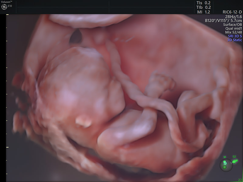
First trimester fetus with Voluson HDlive™
Poon and her colleagues developed a risk assessment based on several biomarkers at 11 to 13 weeks gestation. According to her research, an approach that combines maternal risk factors, uterine artery Doppler, mean arterial pressure, and placental growth factor can identify about 75% of cases of preterm preeclampsia, at a false positive rate of 10%.2-5
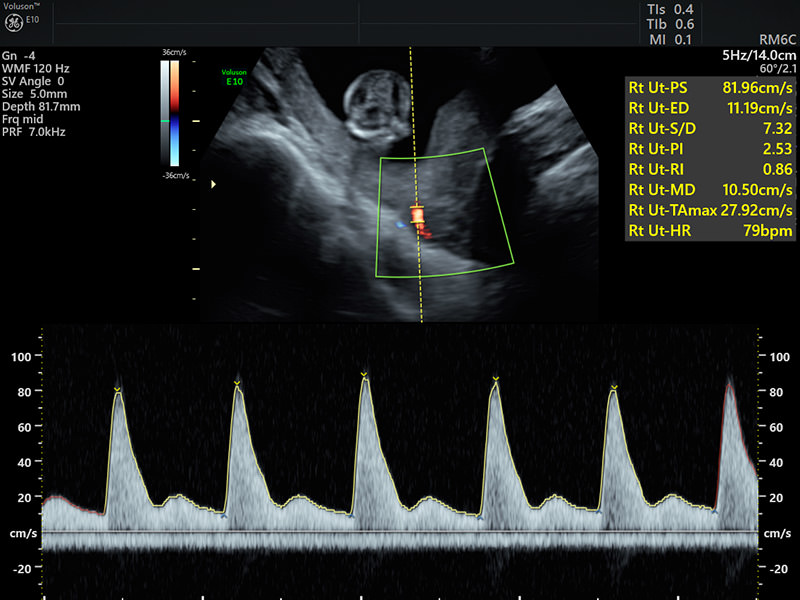
Uterine artery Doppler in the first trimester
For Poon’s method to work, it’s imperative to get accurate ultrasound measurements. Voluson™ from GE Healthcare offers many color Doppler technologies across the platform with excellent sensitivity and spatial resolution. These advancements make it easier to identify the uterine arteries and obtain the necessary measurements to determine the pulsatility index—a key biomarker.
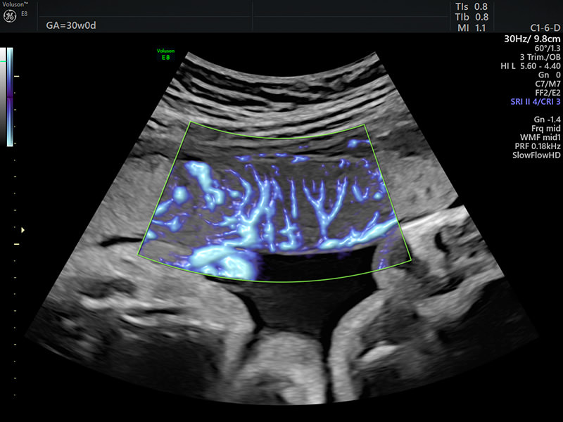
Vascularisation of the placenta with advanced color Doppler technology ‘SlowflowHD’
Once you determine all the biomarkers, the next step is to plug them into an equation. GE Healthcare has made it simple with ViewPoint6TM—intuitive software that allows you to manage your patient information, reports, and images—and directly access the risk assessment algorithm.*
“Many clinicians around the world are using ViewPoint to capture data. If I do a scan, I’ll open ViewPoint, and record the measurement of fetal crown-rump length and fetal nuchal translucency. At the same time, I can enter the data of uterine artery pulsatility index, mean arterial pressure, placental growth factor, and history. I can calculate the risk—all in one program. I think it’s very user-friendly,” Poon said.
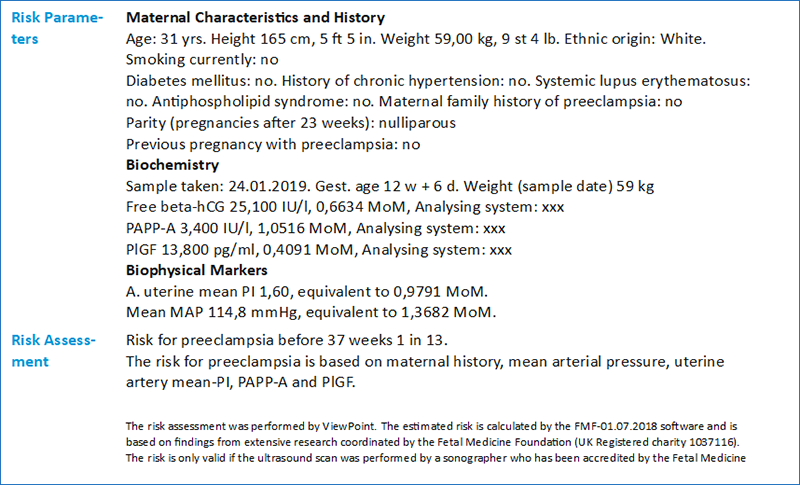
Example of Viewpoint 6 report with the risk assessment of preeclampsia
Private clinicians are slowly adopting the risk assessment program, and it’s expected to gain more momentum with a big endorsement from the International Federation of Gynecology and Obstetrics later this year. But Poon believes it’s going to take a paradigm shift for the program to be implemented universally.
“It's about changing the whole set up of the first trimester scan. You need to have somebody there to take a comprehensive history and it must be accurate because you need to correct the biomarkers for maternal characteristics.” Poon added, “It’s important. We have the potential to substantially reduce the number of women that would otherwise develop preterm preeclampsia. It could be life-changing for all of us.”
*The Preeclampsia Assessment algorithm is not part of ViewPoint6 in some countries.
-
Learn more about Voluson SlowflowHD – a tool which allows you to expand the range of visible blood flow to include low velocities, enabling you to visualize blood perfusion in very small vessels.
-
Learn more about a new standard of color Doppler with Radiantflow – Delivering easy, fast visualization of even the tiniest of vessels.
-
Learn more about Voluson’s HDlive™ imaging technology. While conventional ultrasound rendering uses a fixed light source reflecting light off the skin surface, HDlive™ delivers customizable virtual light sources. Select light target area, direction, and intensity to highlight depth perception and internal structures.
-
Learn more about our Voluson product offerings. Download information about the state-of-the-art clinical innovations offered on the Voluson E10.
-
Learn more about our Voluson product offerings. Download information about how the Voluson S10 Expert can support your everyday routine to complex cases.
-
Learn more about how ViewPoint™ 6 can help streamline your reporting and image management.
World Health Organization. Make every mother and child count. Geneva: World Health Report; 2005.
Rolnik D, Wright D, Poon L. ASPRE trial: performance of screening for preterm pre-eclampsia. Ultrasound Obstet Gynecol; 2017;50:492-495.
Knight M, Kenyon S, Brocklehurst P, Neilson J, Shakespeare J, Kurinczuk JJ, eds. on behalf of MBRRACEUK. Saving lives, improving mothers’ care - lessons learned to inform future maternity care from the UK and Ireland confidential enquiries into maternal deaths and morbidity 2009-12. Oxford: National Perinatal Epidemiology Unit, University of Oxford; 2014.
O’Gorman, Wright D, Syngelaki A, Akolekar R., Wright A, Poon L, Nicolaides K, Competing risks models in screening for preeclampsia by maternal factors at 11-13 weeks gestation. Am J Obstet Gynecol 2016.
O’Gorman N, Nicolaides K, Poon L, The use of ultrasound and other markers for early detection of preeclampsia. Womens Health (Lond) 2016 Mar; 12(2): 199–207.

