Gynecology
- Realize the Possibilities
- Uterine Assessment
- Ovarian Assessment
- Adnexa Assessment
- Pelvic Floor
- Learning Library
Symptoms such as pelvic pain, post-menopausal bleeding, genitourinary dysfunction and infertility, can be confusing and concerning for your patients. They need answers and rely on your expertise for accurate diagnosis, intervention and management. Ultrasound is often the first line of defense in diagnosing gynecological conditions. Voluson™ ultrasound systems deliver exceptional imaging, advanced analysis tools and easy 3D technologies that can provide clinical insights to aid in detection and diagnosis.

Finding Answers
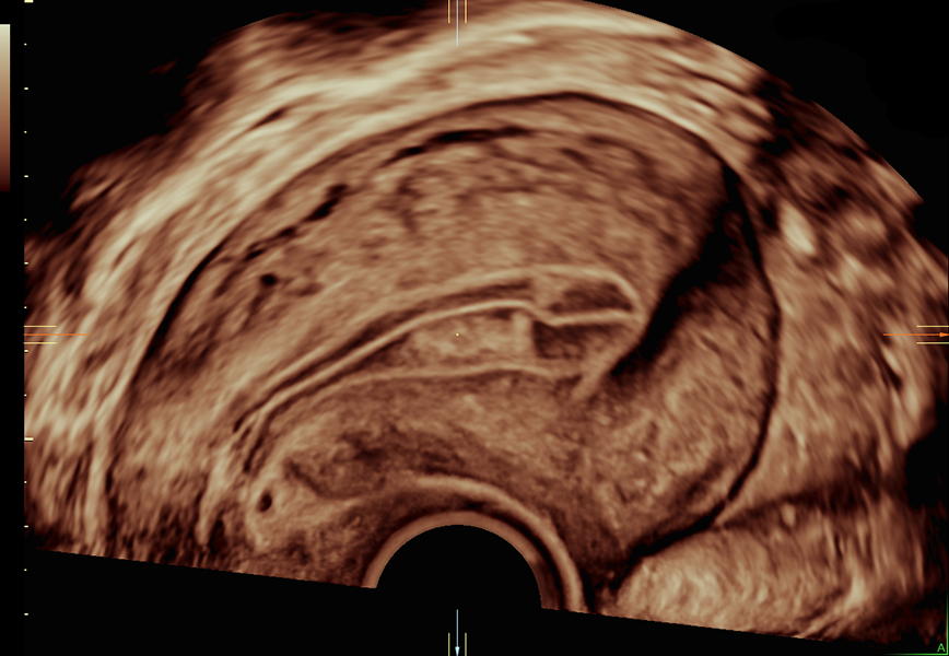
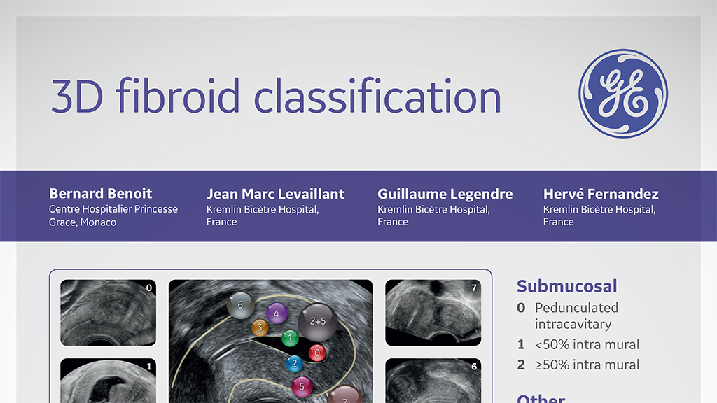
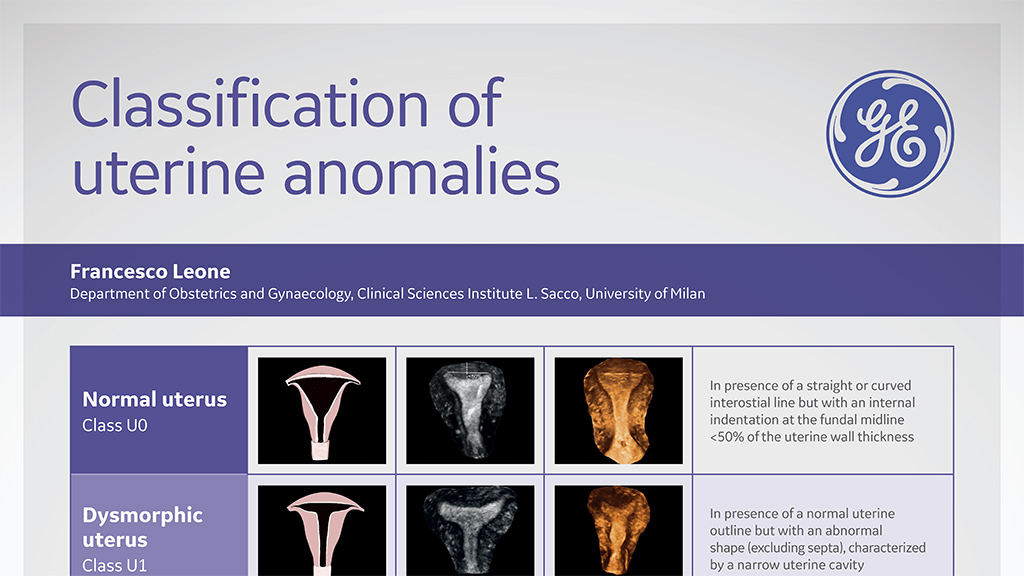
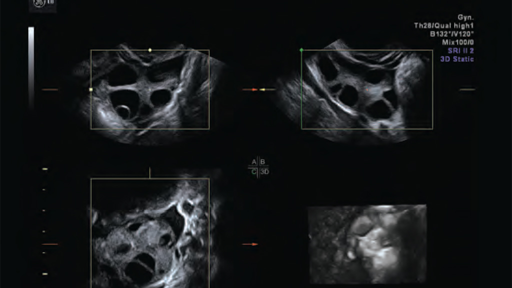
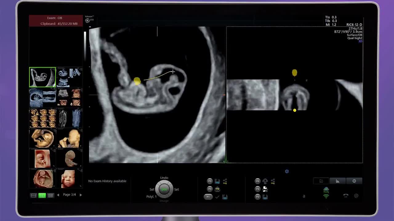
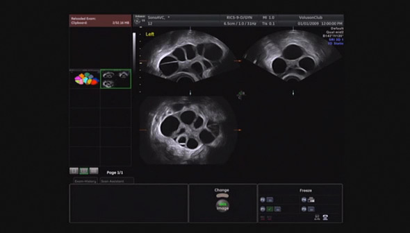
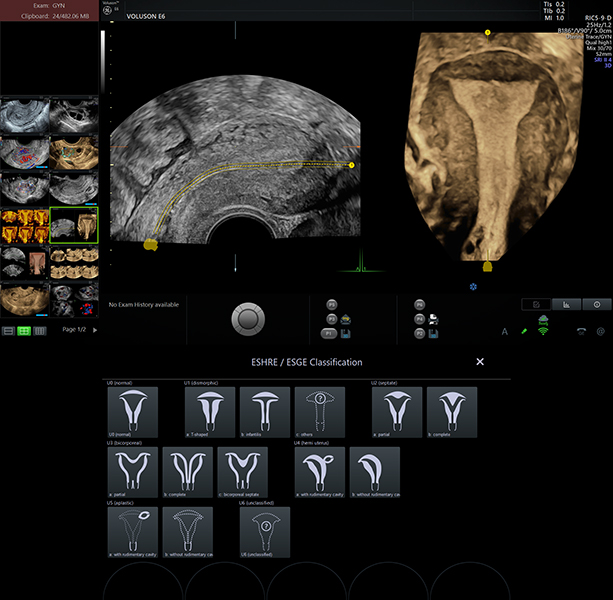
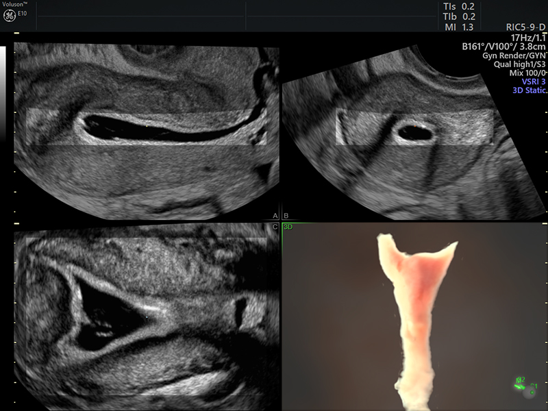
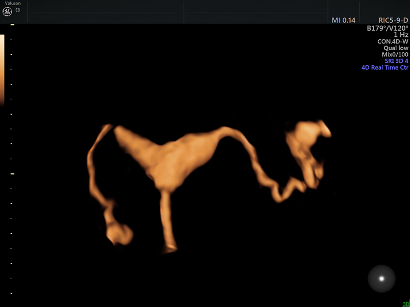
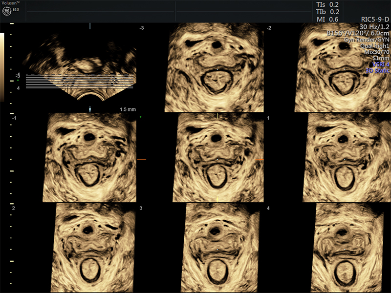
When patients present with pelvic pain or dysfunctional uterine bleeding, endometriosis, adenomyosis or fibroids can be the cause. The ability to easily and rapidly interrogate the uterus and visualize it in any plane using 3D can provide additional information for a more confident diagnosis. In these cases acquiring the coronal plane is particularly helpful. It can also aid in IUCD (Intrauterine Contraceptive Device) localization to determine proper positioning or help to understand the cause of infertility by identifying congenital abnormalities, fibroids or polyps.
CLICK IMAGE in box to scroll through a collection of images.
Simplifying Diagnosis
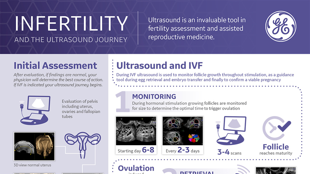
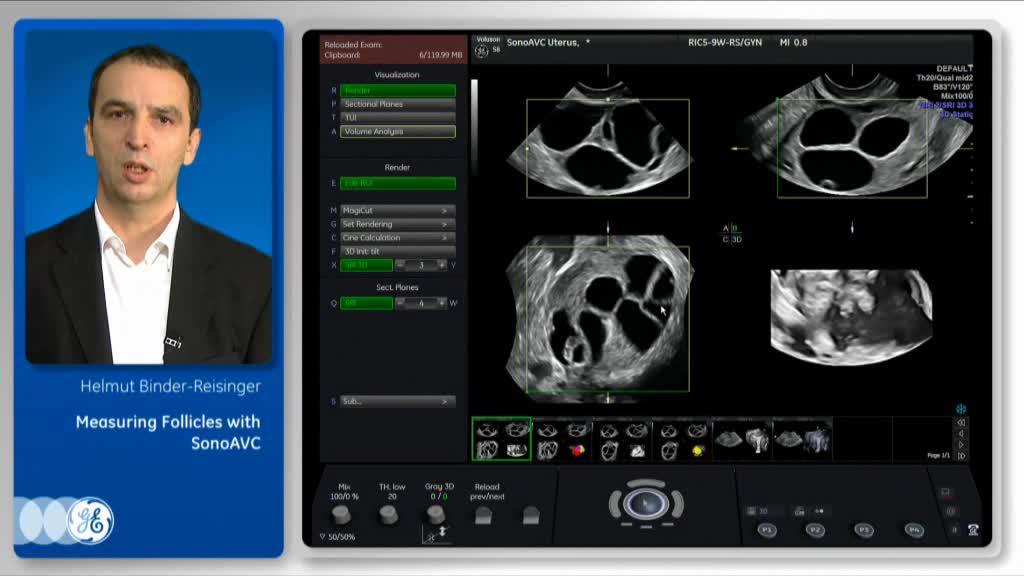
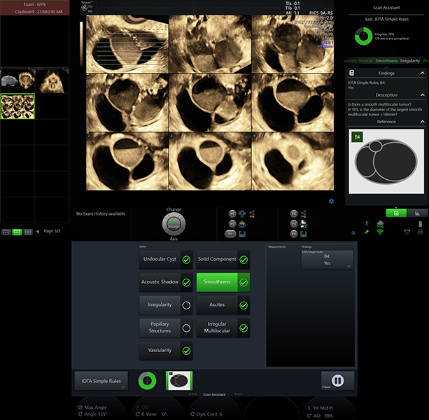
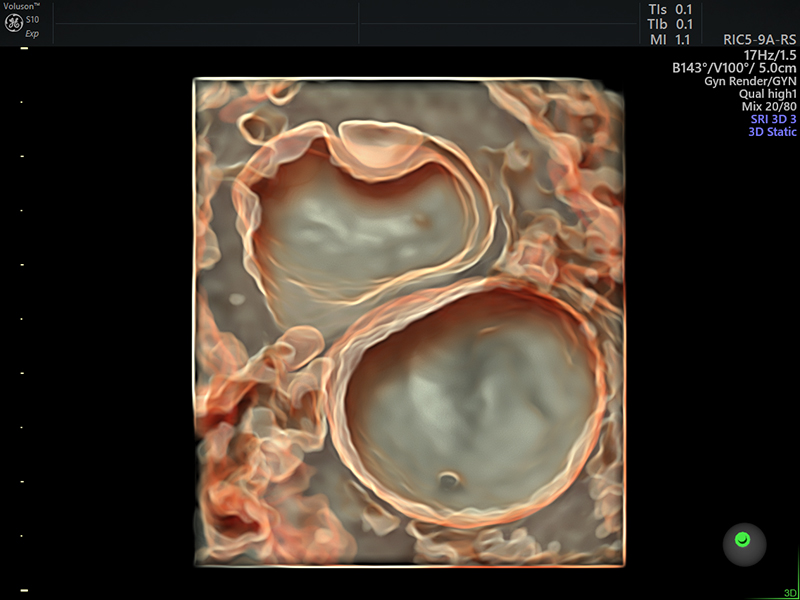
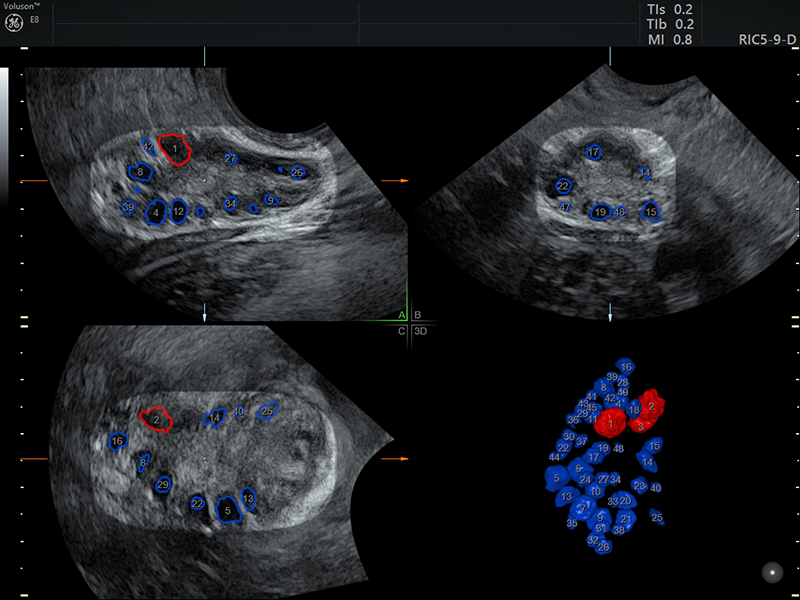
Technologies that efficiently guide you through ovarian assessment can add the information needed for confident diagnoses. Integrated protocols and dedicated reports such as IOTA (International Ovarian Tumor Analysis) and IDEA (International Deep Endometriosis Analysis) evaluate risk for ovarian masses and endometriosis. When assessing the ovaries for infertility the ability to easily capture a 3D volume is essential. SonoAVC™ antral (Sonography-based Automated Volume Count) efficiently counts and categorize follicles and can add important information on ovarian reserve.
CLICK IMAGE in box to scroll through a collection of images.

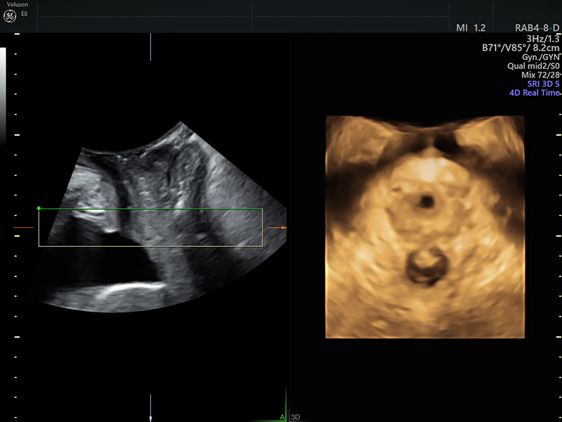
Not Just Uterus and Ovaries
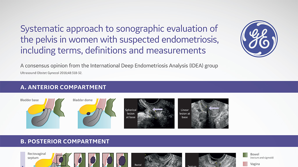
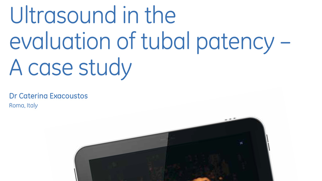
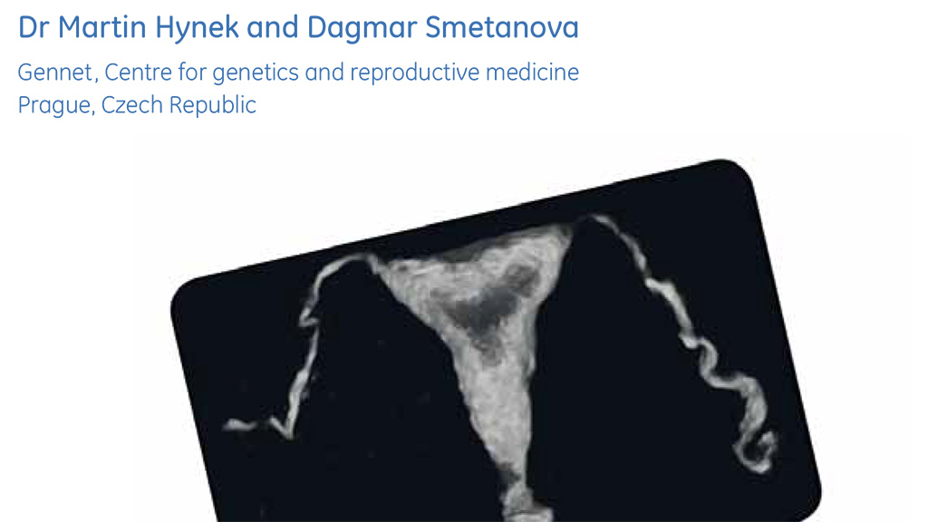
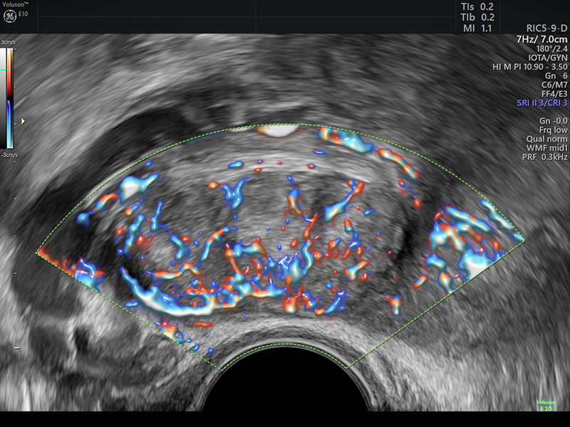
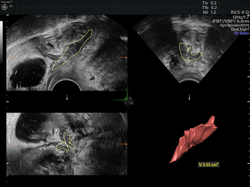
Whether questioning pathology associated with endometriosis, pelvic pain or infertility you are not just limited to just the uterus and ovaries. Understanding complex pathologies and anatomical variants require thorough assessment of the adnexa and spaces adjacent to bladder and rectum, particularly when diagnosing endometriosis. Excellent color Doppler sensitivity is required to understand vascularity of masses, and combined with 3D can enable easier differentiation of hydrosalpinx. Searching for the source of infertility? 3D sonohysterography can help provide information on tubal patency.
CLICK IMAGE in box to scroll through a collection of images.

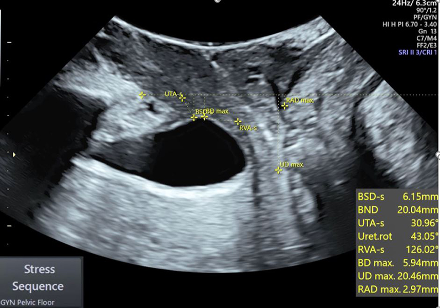
A New Standard of Care
Ultrasound provides real-time diagnostic information which is invaluable in evaluating women with pelvic floor disorders. Pelvic floor ultrasound enables visualization of the pelvic muscles and related anatomy while assessing functionality. It is commonly used to evaluate women with vaginal mesh complications, urinary or fecal incontinence, urinary or sexual dysfunction, pelvic organ prolapse and pain. Efficiently measuring changes to assess functionality is invaluable and the Pelvic Floor Auto-sequencing feature guides you through all of the measurements recommended by the IUGA guidelines.
CLICK IMAGE in box to scroll through a collection of images.


