

During the first trimester of pregnancy, an obstetrician monitors patients for progression, signs of miscarriage and risk of potential problems later on. A first trimester ultrasound provides valuable information to both the obstetrician and patient. Ultrasound images and quality have improved over time, and today's scans reveal much more detail than in the past, allowing for enhanced diagnostic and predictive quality.
First Trimester Considerations
Standard monitoring during the first trimester includes blood tests, genetic tests, ultrasound imaging and physical exams. Obstetricians also look for signs of early pregnancy loss, ectopic pregnancy, problems with placentation, potential risk of miscarriage or preeclampsia, amniotic fluid, birth defects and other risks.
Monitoring allows the physician to address potential issues early on. Birth defects, amniotic fluid levels, placental issues and preeclampsia can increase the risk of stillbirth or pregnancy loss. Ultrasound scans are a reliable way to evaluate risk factors and provide your patient with additional care and monitoring when needed.
Importance of First Trimester Ultrasound
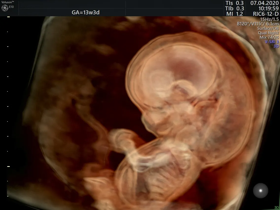
By eight weeks, all the major organ systems are fully developed, but the fetus is only about 1.5 inches in length. An ultrasound after 13 weeks can provide evaluation of birth defects or chromosome abnormalities. In the first trimester, a transvaginal ultrasound provides the best results with high-resolution images. Patients with concerning findings may need additional ultrasounds.
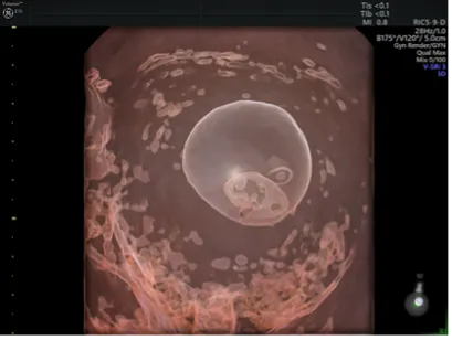
8 week fetus
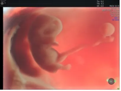
9 week fetus
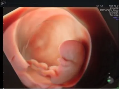
10 week fetus
Pregnancy Confirmation and Location
One of the most basic functions of a first trimester ultrasound is to confirm a pregnancy is intrauterine and viable. Ectopic pregnancy can raise hormone levels in the same way as an intrauterine pregnancy, leading to a positive at-home pregnancy test for the patient. When beta-hCG level measures between 1,500 and 3,000 mIU/mL, an intrauterine pregnancy should be visible on transvaginal ultrasound, according to guidelines published in American Family Physician. An embryo, and embryonic cardiac activity, appears approximately 6.5 weeks after the patient's last menstrual cycle.
Both transvaginal and transabdominal ultrasound are beneficial in evaluating an ectopic pregnancy. Most ectopic pregnancies occur in the Fallopian tubes and require treatment to prevent rupture. Patients should be counseled on other symptoms of ectopic pregnancy, such as dizziness, shoulder pain, uterine pain and bleeding.
Causes of Uterine Bleeding
The American Academy of Family Physicians notes that 25 percent of women experience bleeding during the first trimester of pregnancy. Some bleeding may have a non-obstetric cause, but it may also be a sign of ectopic pregnancy, threatened pregnancy loss or miscarriage. A transvaginal ultrasound can determine the cause of bleeding and provide early intervention.
Early pregnancy loss appears on ultrasound as a gestational sac of 25 mm or greater with no cardiac activity in the embryo. Absence of embryo or cardiac activity less than 85 beats per minute can suggest early pregnancy loss. Bleeding combined with certain ultrasound findings also identifies women at risk of early pregnancy loss. Although the loss may not always be prevented, the physician can closely monitor the patient and provide support where possible.
Ultrasound also allows a physician to spot subchorionic hemorrhage, which when combined with bleeding raises the risk of miscarriage. Repeat ultrasounds may be needed to monitor the pregnancy and assess viability.
Placenta and Amniotic Fluid
First trimester ultrasound also looks for signs of possible placenta previa or other placentation issues. It's often too early to provide an exact diagnosis, but noting the location of the placenta can alert the physician to possible issues.
Amniotic fluid is assessed at every ultrasound using amniotic fluid index (AFI) or single deepest pocket (SDP). Fluid that is too high or too low can lead to poor fetal outcomes. AFI more than 24 cm or SDP more than 8 cm is a sign of polyhydramnios, while AFI less than 5 cm or SDP less than 2 cm is a sign of oligohydramnios, according to a clinical definition published by StatPearls. Ultrasound allows for early detection and intervention of these conditions.
Risk of Preeclampsia
Preeclampsia, which causes high blood pressure after 20 weeks' gestation, impacts between 5 percent and 8 percent of pregnancies, according to the Preeclampsia Foundation. It is a dangerous condition for both the mother and baby. Preeclampsia may be caused by problems with placentation. An increased uterine artery pulsatility index, as measured on Doppler ultrasound, is associated with preeclampsia.
First-trimester uterine artery Doppler combined with biomarker measurements can help identify more patients at risk of developing preeclampsia, according to research in Women's Health. More accurate detection leads to early intervention and proactive management.
Birth Defects and Genetic Syndromes
Certain birth defects can raise the risk of stillbirth or require immediate action after birth. Being able to detect and monitor defects or genetic syndromes provides a higher level of care to the parent and baby.
The first trimester is often too early to identify birth defects, but some structural heart defects may be seen on high-resolution ultrasound images.
Ultrasound also helps clinicians visualize the signs of certain chromosomal defects or genetic syndromes, especially when combined with blood tests. When undergoing IVF, a first trimester ultrasound in conjunction with preimplantation genetic testing is effective at looking for aneuploidy, particularly trisomy 21 (also known as Down syndrome). A nuchal translucency measurement with a cut-off of 3.5 mm has the highest sensitivity and specificity to detect congenital abnormalities, according to research published in Prenatal Diagnosis.
An ultrasound in the first trimester can provide valuable information for a pregnancy—whether confirming a healthy, viable pregnancy or helping identify potential risks for mother and baby. Early detection with specialized ultrasound scans gives both the physician and patient the chance to intervene early or plan for a course of action at birth.
Cystic Hygroma seen at 12-weeks

