Image Quality
2D image quality enhancement tools
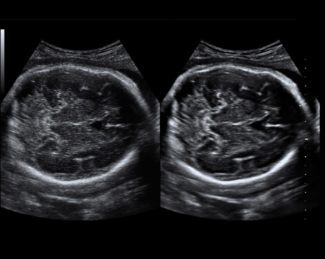
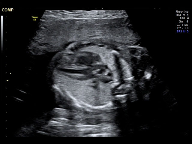
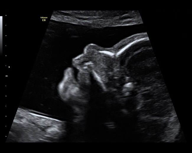
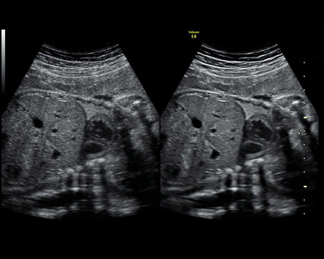
- Advanced Speckle Reduction Imaging (SRI) — helps heighten the visibility of organs and lesions with high-definition contrast resolution that suppresses speckle artifact while maintaining true tissue architecture.
- CrossXBeamCRI* — helps enhance tissue and border differentiation with an innovative, real-time spatial compounding acquisition and processing technique.
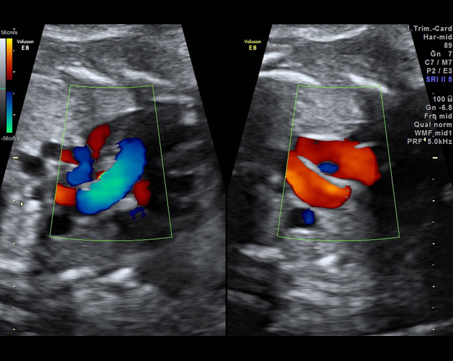
- HD-Flow* — uses a bi-directional Doppler feature to help achieve a sensitive vascular study and helps reduce overwriting.
- Focus and Frequency Composite (FFC) — provides for both penetration and resolution benefits by allowing simultaneous transmit frequencies — high frequencies in the near-field together with low frequencies in the far-field.
- Coded excitation — uses GE’s innovative coded technology to allow high frequencies to penetrate deep into the body for exellent resolution on technically difficult patients.
- HD-Zoom – helps users localize regions of interest for closer study.
- Pulse inversion (harmonics) — utilizes 2 pulses to create an image, each an inverse of the other, cancelling out noise to help produce a clean image.
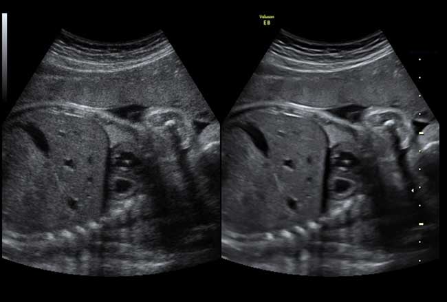
- Dual-view enables simultaneous visualization of anatomy and blood flow.
- Wide sector for endocavitary probes – enables more anatomical information to be displayed in a single image versus the standard field of view.
Volume Imaging
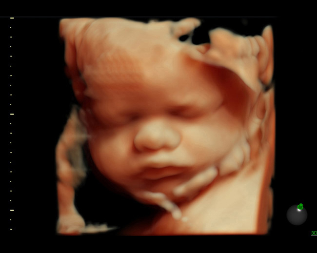
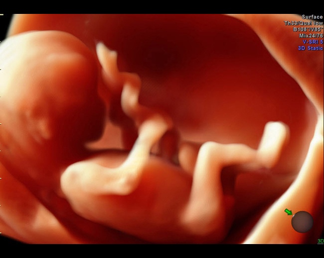
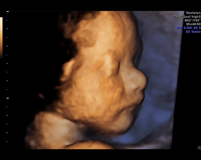
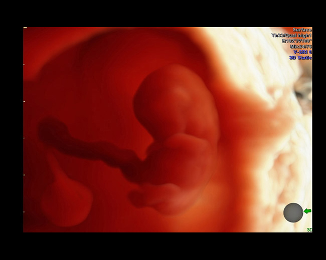
- HDlive — This innovative rendering tool provides exceptional anatomical realism and helps increase depth perception. This imaging capability can help you achieve a deeper understanding of relational anatomy, enrich patient communication and help enhance diagnostic confidence.
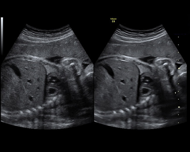
- Volume SRI (V-SRI) — This tool provides a high level of speckle reduction utilizing volume/voxel versus traditional single slice imaging. It helps improve 3D/4D quality in multi-planar studies and rendered mode, and also provides an enhanced smoothing effect on rendered images which can help improve diagnostic confidence.
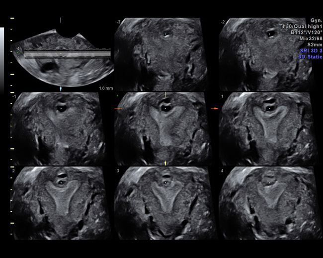
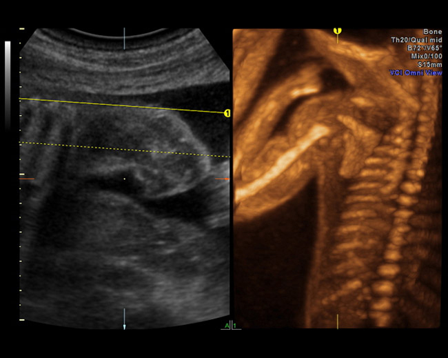
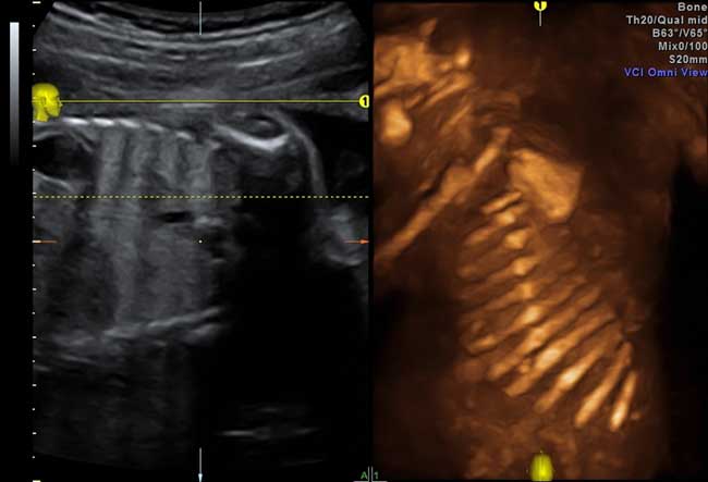
- Advanced Volume Contrast Imaging (VCI) with OmniView — Diagnostic confidence in sonography requires the ability to differentiate irregular shapes with precision. This tool can help improve contrast resolution and visualization of the rendered anatomy with clarity in any image plane.
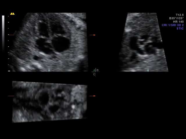
- STIC (Spatio-Temporal Image Correlation) — A technology that was pioneered by Voluson engineers. STIC captures a full virtual fetal heart cycle in real time, and the volume can be saved for off-line analysis.
- Advanced 4D including TUI (Tomographic Ultrasound Imaging) — This tool combines advanced 4D imaging capabilities with TUI (a simultaneous view of multiple parallel slices of a volume data set) which can help you make analysis and documentation of dynamic studies easier.
Advanced Probe Technology
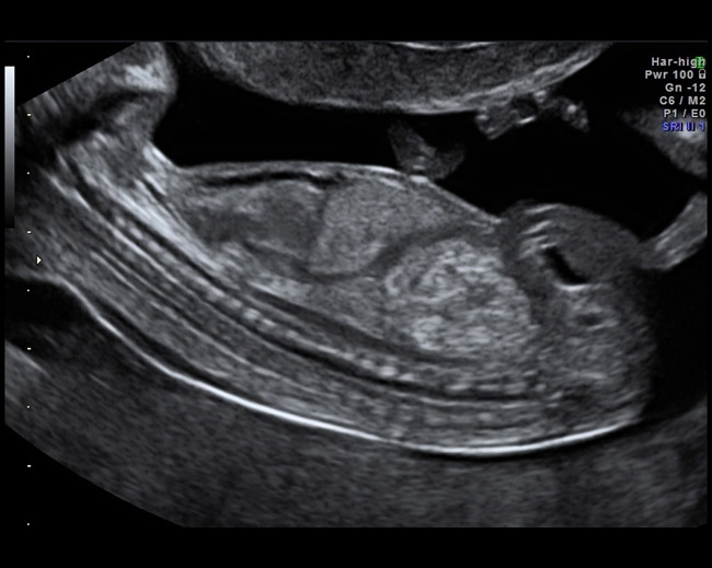
View images created with advanced Voluson probes and demonstrate extraordinary image quality.
- SonoRender Start — enables the user to quickly optimize volume rendering with the touch of a single button.
- Beta View — steer the scan plane to any oblique direction, helps reduce probe manipulation and increase patient comfort.
- Wide variety of rendering techniques – Surface, Skeleton, Inversion, Glass Body; all designed to help enable you to see what you need within a volume.
- Intuitive user interface – Consistent, easy to operate and 3D/4D volume controls.
Probe Technology
The Voluson systems supports a wide range of advanced 2D and 3D probes, which enable high quality images with exceptional reliability. System intelligence and probe technology combine to produce outstanding image quality with optimized settings to help minimize user interations – just place the probe and scan with confidence.
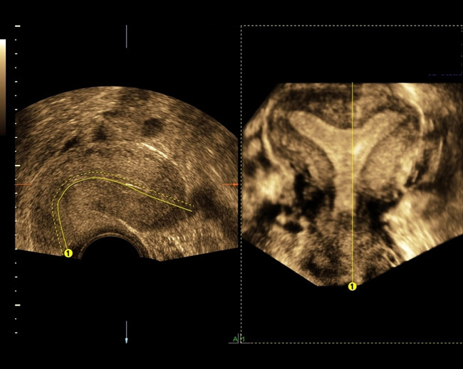
- RIC6-12-D — this high resolution endovaginal volume probe helps you detect fine details early in the first trimester and in gynecology exams.
- New RAB6-D — fatigue can be a thing of the past with this innovative volume probe that is 40% lighter than the previous version. Its ergonomic design and outstanding image quality in 2D and 3D/4D, features a thin flexible cable and sits comfortably in the operator's hand.
- C1-5-D — this probe helps you image confidently with deep penetration even when scanning large patients.
- New C4-8-D — with high frequency and a small footprint, this probe helps provide exceptional high resolution obstetrical images during each trimester.
- RM6c — this high-resolution convex matrix technology volume probe provides excellent spatial resolution and image uniformity from near to far field.
- 9L-D — this linear probe helps provide high-quality images in the first trimester.
Advanced Probe Technology
View images created with advanced Voluson probes and demonstrate extraordinary image quality.
- ML6-15-D — this linear probe features matrix technology for breast imaging, helping provide maximum spatial resolution and image uniformity in a 50mm footprint.
- RIC5-9-D transvaginal — equipped with wide sector technology (2D) and 120° volume angle (3D/4D), enabling you to see more anatomical information displayed in a single image or volume.
- 11L-D linear — high frequency linear transducer designed for breast and other small parts imaging.
- 3Sp-D-sector probe to help you with your shared service imaging needs. Adding cardiac imaging capabilities can help expand the use of your Voluson.
Please contact your Regional sales representative for probe configurations per Voluson product.
Automation Tools
Voluson features advanced tools to help improve reproducibility and efficiency of obstetric and gynaecologic exams, including:
- SonoNT* (Sonography-based Nuchal Translucency) and SonoIT* (Sonography-based Intracranial Translucency) – Voluson technologies that help provide semi-automatic, standardized measurements of the nuchal and intracranial translucency as early as 11 weeks. Both tools can integrate easily into your workflow. SonoNT helps reduce the inter-and-intra-observer variability that comes with manual measurements, and helps provide you with the reproducibility you demand.
- SonoBiometry* – Performs a semi-automatic measurement of the head (both head circumference and bi-parietal diameter), abdomen and femur. This tool can help enhance clinical workflow through helping reduce keystrokes to perform biometry measurements.
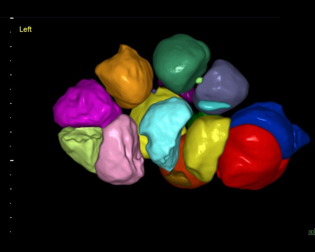
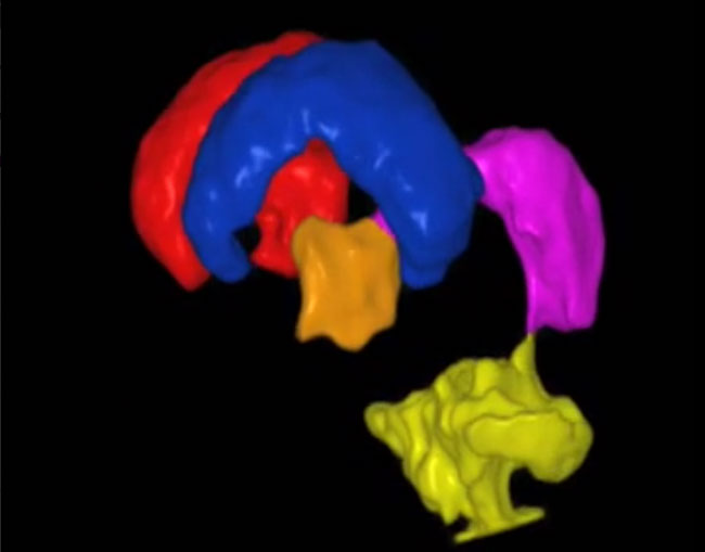
- SonoAVC* general (Sonography-based Automatic Volume Count general)
- SonoAVC* follicle (Sonography-based Automatic Volume Count follicle) — semi-automatically calculates the number and volume of hypoechoic structures from a 3D ovarian volume. Helps improve efficiency and workflow of follicular assessment.
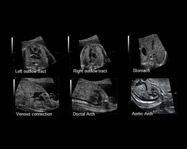
- SonoVCAD* heart (Sonography-based Volume Computer Aided Display heart) — assists in generating views of a fetal heart from a four-chamber view and complies with the recommended AIUM standard examination of the fetal heart.
Advanced Probe Technology
View images created with advanced Voluson probes and demonstrate extraordinary image quality.
- SonoVCAD* labor (Sonography-based Volume Computer Aided Display labor) — helps you to measure fetal head progression, rotation and direction while automatically documenting the labor procedure with objective ultrasound and manual data in one easy report.
- STIC (Spatio-Temporal Imaging Correlation) captures a full fetal heart cycle beating in real time, and the volume can be saved for offline analysis.
- SonoRender Start enables the user to quickly optimize volume rendering with the touch of a single button.
Exam consistency and Quality
- Scan Assistant — by automating many repetitive actions that occur during an exam, this tool helps improve quality assurance, exam consistency, and productivity.
- Virtual Rescan — the system captures raw data with every exam, enabling a virtual rescan of the patient at any time after the study, on the system or at a remote workstation such as ViewPoint*.
Streamline archiving and reporting
- DICOM** — compliant and compatible with many electronic medical record systems
- ViewPoint — reporting and image management enables clinicians to create electronic reports that include images and charts, enhancing archiving and physician communication. ViewPoint also enables volume manipulation with 4D View option, and ultrasound data importation to your medical record system.
