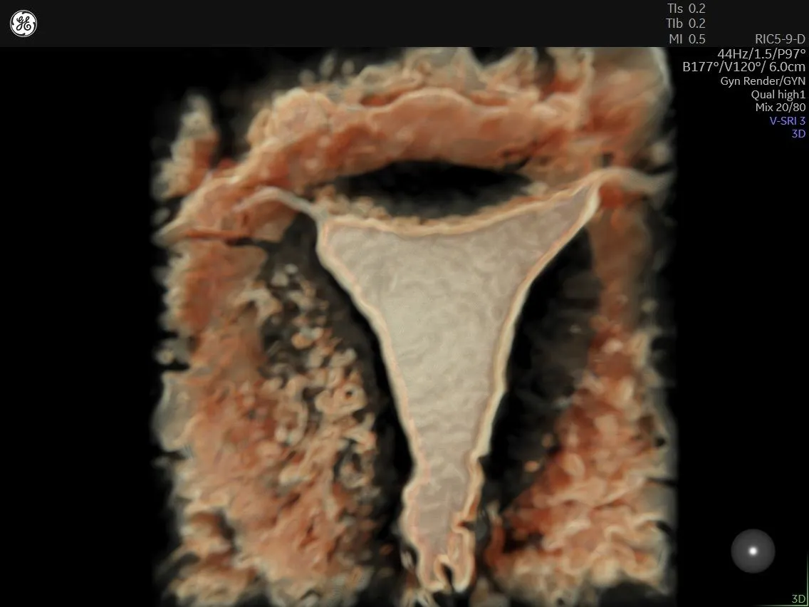
A Guide for Advancements in 3D Ultrasound: Enhancing Gynecological Diagnosis and Imaging
Ultrasound has numerous uses in gynecology, from monitoring a fetus during pregnancy to examining the uterus and ovaries. One of the most significant advancements in ultrasound is 3D ultrasound, which began gaining steam in the mid-1990s with the production of the 530D Voluson™.
One of the major differences between 2D and 3D ultrasound is that 2D ultrasound only allows sonographers to acquire a single section of an area of interest, says Dr. Mala Sibal, a consultant physician at the Department of Fetal Medicine and Obstetrics and Gynecology Ultrasound at Manipal Hospital in Bangalore. Sibal is also the director of Gynecology Academy, an online learning portal for gynecological ultrasound and fetal and maternal medicine.
With 2D ultrasound, the display is fixed, and there is no scope for getting more information post-acquisition. It is completely operator-dependent. On the other hand, 3D imaging can acquire an entire volume, which physicians can then study in various display formats and planes. It is also relatively operator-independent, and sonographers can work on volumes later to get more information.
Steps for 3D Ultrasound
The first step for 3D ultrasound is data acquisition, which starts with good 2D views, Sibal says. The next step is to choose the display mode, of which there are several:
- Multiplanar display. This allows the user to visualize the area of interest in three dimensions simultaneously
- Tomographic display. This display shows parallel sections of the volume, similar to CT and MRI
- Surface rendering. This allows the user to view any surface or plane that is of interest; it is the most commonly used display modality in gynecological ultrasound
- Omniview. This mode is useful for uterine anomalies. Omniview allows the user to draw a polyline along the plane of interest; it provides a coronal plane of the cervix and uterus in a single image
- Glass body display. This display combines surface or transparent rendering with Doppler, and it provides a transparent mode that allows the user to see the background cervix and uterine body
- Volume analysis. This allows the user to draw around the region of interest in various dimensions and get an accurate volume
These display modes are available as presets on ultrasound machines, and users can switch back and forth between display formats at any time.
After choosing your display mode, you can go into volume and imaging processing, Sibal says. These modalities include volume contrast imaging (VCI), electronic scalpel, contrast and brightness control, color selection, and edit light.
After processing the images, the sonographer can store them. This is another advantage over 2D, Sibal says. With 3D, you can work on the volume at a later time, which can be beneficial in situations where the clinic is busy or the patient is in pain. It can also be useful for sharing images with colleagues for a second opinion or for research studies.
Uses of 3D Ultrasound in Gynecology
Of the several uses of 3D ultrasound in gynecology, one is routine ultrasound. "Some people use 3D as a routine in their pelvic scan," Sibal says. "We know we can get very good images of the uterus on long section and transverse section, both on transabdominal and transvaginal scans." While it is impossible to get a coronal section of the uterus with 2D, 3D makes it possible.
There are also certain conditions where a diagnosis cannot be made without 3D, such as with uterine anomalies or pelvic floor studies. Visualizing the position of an intrauterine contraceptive device (IUCD) is also made possible with 3D. While 2D can give the suspicion of a uterine anomaly, 3D is necessary to confirm its presence and classify the anomaly because it gives the contour of the uterine fundus and the shape of the uterine cavity. If the contour of the uterine fundus is abnormal, based on the new American Society for Reproductive Medicine (ASRM) classification, and if the indentation is more than 10 millimeters, this indicates either a bicoordinated uterus or diadelphous uterus.
"If the contour of the uterine fundus is normal, the next thing we need to look at is the shape of the endometrial cavity, which may be normal or abnormal," Sibal says. "If the indentation is less than 10 millimeters, we call it an arcuate uterus. If it is more than 10 millimeters, we call it a separate or a partial septate uterus."
With pelvic flow studies, sonographers need to get a coronal section image of the pelvic floor to look for symmetry, which can't be done with 2D. Here, 3D helps with locating IUCDs. It provides a coronal rendered image of the uterine cavity and can produce a single image showing the endometrium and IUCD distinctly.
Another area where 3D can serve as a value add for 2D ultrasound is fibroid mapping. Although 3D isn't necessary for every patient with fibroids, it is essential if there is a fibroid adjoining the endometrial cavity. It allows the sonographer to see exactly how the fibroid is distorting the shape of the endometrial cavity and can help confirm if the fibroid is completely within the cavity.
3D is also beneficial in areas such as determining antral follicle counts and measuring the volume of stroma compared with cystic areas, which is useful for studies on polycystic ovaries. Intrauterine additions can also be detected with 3D imaging using coronal rendering.
Sibal notes a case where a patient came in for a nuchal translucency scan at 12 weeks, five days. She noticed a cyst that had ground-glass contents, but there was a lot of solid tissue that she couldn't see the margins of. She couldn't tell if it was a malignancy or a decidualized endometriotic cyst. She decided to do surface rendering, which helped her delineate the margins of papillae that were not clear with 2D imaging.
"When you have challenges, you should use multiple modalities, and 3D is one such great modality to use," Sibal says. "3D offers a vantage point one just cannot miss doing gynecological ultrasound. It displays views that provide or highlight additional features that are not appreciated on 2D imaging." Knowing which display formats to use and which image processing to apply can go a long way toward making 3D imaging an essential tool for gynecological ultrasound.
View Dr. Maga Sibal's full video – The importance of 3D ultrasound in gynecology.

