

Ultrasound is making headway as the imaging tool of choice for evaluating adnexal masses. As a result, many clinical organizations are working to standardize adnexal mass ultrasound terms and meanings, especially for abnormal adnexa ultrasound. But why does it sometimes feel like clinicians are speaking different languages when it comes to reporting on these masses?
Currently, several lexicons are used to describe adnexal masses in North America. Two common systems are the International Ovarian Tumour Analysis (IOTA) guidelines and the Ovarian-Adnexal Reporting and Data System (O-RADS). The goals for both systems are to improve communication, reduce ambiguity, predict the risk of malignancy and formulate evidence-based algorithms for clinical management.
Common Adnexal Mass Ultrasound Terms and Meanings
A common language for describing adnexal masses is essential. Clinicians such as pediatricians, emergency physicians, radiologists and oncologists all rely on ultrasound reports while counseling their patients and assessing treatment options.
Using clear and standardized language can also benefit your patients. Knowing when a mass is normal or abnormal at an early stage reduces the number of unnecessary surgeries and helps triage patients to other care centers for appropriate treatment.
Even common terms should be examined to make sure we're all speaking the same language. The journal Radiology suggests that the term "follicle" should replace "cyst," for instance, given that "cyst" indicates disease in most organs but refers to a normal, fluid-filled structure in the ovaries.
IOTA Guidelines
The European IOTA Group first published its new list of standardized terms for adnexal lesions in 2000. The word "lesion" was deliberately chosen to imply an abnormality. This consensus opinion allowed a new era of research using B-mode imaging and color Doppler examination of adnexal lesions to blossom.
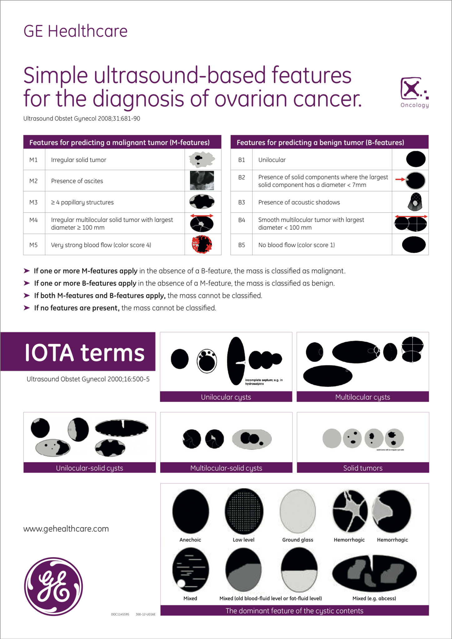
Before the IOTA guidelines, previous adnexal lesion studies had reached differing conclusions using similar algorithms — in part because of differences in the terminology they used. IOTA identified six categories of adnexal lesions based on a qualitative description of their solidity and number of chambers. These categories apply to both the ovaries and the fallopian tubes. IOTA went on to define morphologic features ranging from papillary projections to the presence of fluid in the pouch of Douglas.
After standardizing clinical terminology throughout 24 centers in 10 countries and collecting ultrasound data from almost 6,000 women, IOTA developed the "Simple Rules" and ADNEX model. These models predict a lesion's cancer potential, from benign to metastatic, and are now a standard tool onboard some ultrasound machines.
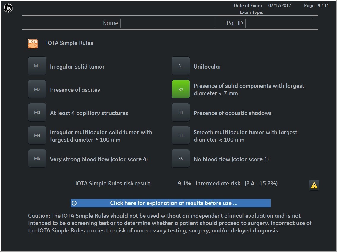
IOTA simple rules on Voluson E10 system for ultrasound terms and meanings
IOTA ultrasound terms and meanings
O-RADS Guidelines
One alternative to the IOTA guidelines is the American College of Radiology (ACR) O-RADS lexicon, developed in 2015. The ACR argues that adoption of the IOTA models has been limited in North America, giving rise to alternatives. O-RADS was designed as a universal vocabulary to improve communication between doctors doing the imaging and doctors applying the results in a clinical situation.
A key part of creating a common lexicon, the ACR argues, is retiring terms that may be vague or misleading to patients. One of the previously common descriptors that O-RADS discourages, for example, is "complex."
Who Uses Adnexal Mass Ultrasound Guidelines?
In North America, sonographers perform the majority of pelvic ultrasounds, with radiologists, gynecologists and physicians reviewing the images and dictating the final report. The American Institute of Ultrasound in Medicine offers voluntary certification to these practitioners demonstrating they have met national standards in ultrasound, but focuses on requiring measurements and rigorous documentation of abnormal adnexa ultrasound instead of offering its own list of terms.
A common medical language is key for avoiding misunderstandings and confusion. Learning a common ultrasound lexicon can help providers be a part of a unified conversation about gynecological health.

