Advancing Superficial Endometriosis Detection Through High-Frequency Ultrasound
Superficial endometriosis is the most common form of the condition, comprising nearly 80% of endometriosis diagnoses.1 However, the condition has historically evaded ultrasound detection due to the shallow nature of the lesions, which places them at the edge of conventional transvaginal ultrasound resolution, leading to incomplete preoperative disease mapping and delayed diagnoses.
Laparoscopy has been considered the gold standard for diagnosing endometriosis as it allows direct visualization of endometriotic lesions, but national and international guidelines have shifted focus to non-invasive imaging-based diagnosis.2 In response, the International Deep Endometriosis Analysis (IDEA) consensus opinion has standardized ultrasound examination of patients with endometriosis.
Understanding the IDEA Consensus Amendment
The original 2016 IDEA consensus established a standardized four-step transvaginal ultrasound protocol for diagnosing and mapping endometriosis.3 The protocol created a reproducible approach for identifying and mapping deep infiltrating endometriosis (DIE) and communicating the findings across multidisciplinary care teams.
The 2022 guidelines set by the European Society of Human Reproduction and Embryology (ESHRE) reinforced ultrasound and magnetic resonance imaging (MRI) as first-line diagnostic tools for endometriosis, while also acknowledging the limitations in detecting superficial peritoneal lesions.4 That acknowledgment created a pathway for further refinement of the diagnostic framework.
The 2025 IDEA addendum closed the gap by expanding the framework to include structured assessment of superficial endometriosis. The addendum introduces standardized terminology for peritoneal implants, criteria for lesion characterization, and guidance on integrating superficial findings into existing reporting structures. Early, non-invasive identification supports more precise surgical planning, guides fertility preservation decisions, and has the potential to reduce diagnostic delay.
The Physics Behind High-Frequency Probe Superiority
Higher-frequency transducers, which typically operate between 9 and 15 MHz, produce shorter wavelengths that yield high-resolution images of superficial structures. As frequency increases, depth penetration decreases, but because superficial endometriotic lesions lie within the transducer's near field, the trade-off works in the clinician’s favor.
High-frequency imaging is particularly valuable when evaluating:
- The anterior pelvic compartment
- The bladder peritoneum
- The uterosacral ligaments
- The rectovaginal septum
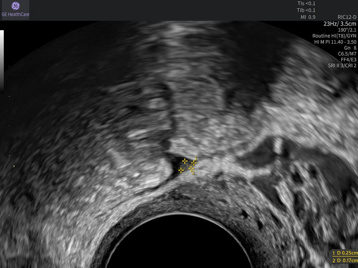
On high-frequency ultrasound, superficial endometriotic lesions may appear as subtle, hyperechoic or hypoechoic projections from the peritoneal surface measuring less than 5mm.5 They may also appear as filmy adhesions, cystic areas, or peritoneal pockets. Clinicians must optimize their probe selection and scanning techniques to maximize diagnostic yield.
Scanning Protocols for Superficial Endometriosis Detection
Patient preparation optimizes visualization. A moderately filled bladder improves anterior compartment assessment, while bowel preparation reduces acoustic shadowing in the posterior compartment when indicated.
The systematic peritoneal examination follows a structured sequence aligned with the IDEA framework, evaluating the anterior, central, posterior, and lateral compartments along the peritoneal surface and organ serosa.
The transvaginal probe should be positioned within the posterior fornix in the midsagittal plane without compressing the Pouch of Douglas (POD), preserving a small pocket of fluid for peritoneal visualization. Push-pull maneuvers with the transducer redistribute fluid posteriorly, while manual compression moves fluid laterally to evaluate ovarian fossae.
The sliding sign adds a functional dimension to morphological assessment. A negative sliding sign suggests posterior compartment adhesions and warrants increased scrutiny for peritoneal involvement.
With the probe placed in the anterior fornix, superficial endometriosis can be visualized if there is fluid in the anterior vesicouterine pouch. Scanning should continue slowly in the sagittal, axial, and transverse planes. Scanning tangentially to the peritoneum helps visualize superficial lesions in all compartments.
Bridging the Gap Between Equipment and Expertise
The most advanced high-frequency transducer can't replace thoughtful image interpretation, anatomical knowledge, or clinical judgment.
Diagnostic confidence in superficial endometriosis detection is built through ongoing, structured, practice-embedded education, not just equipment acquisition. Case-based learning aids skill development in complex gynecological imaging,6 helping clinicians translate evolving consensus recommendations into routine clinical practice. Reviewing diverse clinical cases sharpens pattern recognition for subtle lesion appearances. Engaging in peer review also helps reinforce best practices and uncover possible scanning errors.
GE HealthCare’s (GEHC) commitment to clinician empowerment reflects this understanding. Through structured education programs designed for gynecological imaging specialists, GEHC supports practitioners in moving from equipment familiarity to diagnostic mastery, providing a clinical education infrastructure that complements consensus statements.
Leading the Next Era of Endometriosis Imaging
The IDEA consensus amendment marks a watershed moment in the evolution of endometriosis imaging. Incorporating superficial endometriosis into standardized ultrasound assessment expands opportunities for comprehensive disease detection and improved surgical planning. While high-frequency ultrasound technology provides the image quality needed to visualize superficial endometriosis, the clinician remains the diagnostic authority.
As comprehensive endometriosis assessment becomes the expected standard of care, clinicians who combine updated consensus guidance with advanced image interpretation and access to continuous clinical education will be better equipped to deliver accurate diagnoses, informed treatment planning, and improved outcomes for women living with the condition. GEHC is committed to supporting that journey, from the physics of probe selection to learning environments that sustain diagnostic confidence over time.
Explore GEHC’s gynecological education resources to deepen your superficial endometriosis detection skills and align with the expanded IDEA framework.
Interested in Learning More?
Explore expert-led education on ultrasound assessment of endometriosis:
Dr. M. Leonardi: Mastering ultrasound for superficial endometriosis detection.
ISUOG: Endometriosis uncovered: mastering ultrasound for excellence
Dr. S. Johnson: How to diagnose endometriosis using ultrasound
Professor G. Condous: Ultrasound imaging of endometriosis
References:
1.International Society of Ultrasound in Obstetrics and Gynecology. Superficial endometriosis. ISUOG. May 2022. Accessed July 16, 2026. https://www.isuog.org/clinical-resources/patient-information-series/patient-information-gynecological-conditions/superficial-endometriosis.html
2.Guerriero S, Condous G, Rolla M, et al. Addendum to consensus opinion from the International Deep Endometriosis Analysis (IDEA) group: sonographic evaluation of superficial endometriosis. Ultrasound Obstet Gynecol. 2025;66(4):541-547. doi:10.1002/uog.29288
3.Guerriero S, Condous G, van den Bosch T, et al. Systematic approach to sonographic evaluation of the pelvis in women with suspected endometriosis, including terms, definitions and measurements: a consensus opinion from the International Deep Endometriosis Analysis (IDEA) group. Ultrasound Obstet Gynecol. 2016;48(3):318-332. doi:10.1002/uog.15955
4.Becker CM, Bokor A, Heikinheimo O, et al. ESHRE guideline: endometriosis. Hum Reprod Open. 2022;2022(2):hoac009. doi:10.1093/hropen/hoac009
5.Pedrassani M, Guerriero S, Pascual MA, et al. Superficial endometriosis at ultrasound examination—a diagnostic criteria proposal. Diagnostics. 2023;13(11):1876. doi:10.3390/diagnostics13111876
6.Zhu Y, Zhang J, Fei J, Fang H, Zhang Z. Problem-based learning and case-based learning in clinical practical teaching for gynecology residents: a narrative review. Adv Med Educ Pract. 2025;16:1269–1279. doi:10.2147/amep.s534053

