Applications of the SonoCNS Tool for Prenatal Imaging: Doctors' Perspectives
Mid-trimester prenatal imaging is the primary tool to monitor for brain anomalies in the developing fetus. Recommended practice guidelines for fetal brain imaging from the International Society of Ultrasound in Obstetrics and Gynecology (ISUOG) call for this assessment. Yet, manual scanning may consume precious minutes in a prenatal care visit or depend on the skill of the operator. The AI-assisted SonoCNS tool is a modern solution to address challenges in fetal brain imaging, offering accurate, reproducible fetal brain measurements that today's pace demands.
The Future Is Now in AI-Assisted Imaging
The advent of artificial intelligence has improved clinical efficiency and analysis in many corners of women's health. In ultrasound assessment, AI-assisted tools can simplify routine scans to aid in clinical decision-making. Now, the innovative SonoCNS harnesses the power of AI for brain imaging of the fetal patient, promising to streamline clinical workflows and improve the detection of normal brain development and abnormalities during the prenatal care visit.
In today's clinical practices, time is one of the most significant factors relating to care. Traditional methods of capturing measurements of the fetal brain require a long list of procedural steps, consuming valuable time with the prenatal patient. The task of manipulating volume data to find specific planes to investigate fetal anatomy also demands precision and skill. With traditional imaging systems, the skill and experience of the sonographer may introduce variability and influence results.
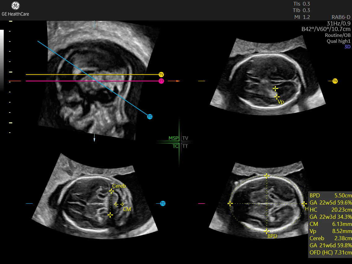
SonoCNS is an AI-assisted tool that provides reliability, reproducibility, and accuracy. A few simple taps by the physician or sonographer is all that's needed to obtain a volume of the fetal head within seconds. Then, SonoCNS identifies the appropriate planes and performs the measurements. Tomographic display of the fetal brain volume easily illuminates structures, allowing physicians to confirm normality or identify fetal brain malformations during the prenatal exam.
SonoCNS provides greater speed—without sacrificing clinical reliability or accuracy of prenatal imaging. The new generation of SonoCNS is now 29 percent faster than an earlier version.
Time Efficiency and Automation
The Voluson™ ultrasound platforms embeds intelligence in fetal imaging with the innovative SonoCNS system, revolutionizing the routine assessment of fetal brain structures. In an era where time is of the essence, the integration of these twin technologies shortens the period spent gathering the three standard planes and five recommended measurements through an alternative automated workflow.
The workflow begins with the physician or sonographer acquiring a 3D volume of the fetal head, starting from or near the transthalamic plane position. SonoCNS then proposes the three required planes in the fetal brain. The display shows their position in relation to a presentation of the mid-sagittal plane. At the press of a button, the AI-assisted tool performs the five required measurements. Control remains with the user, who can add all results directly to the report or adjust any individual caliper beforehand, either in the multiplanar or full-screen displays.
The device is so intuitive and easy to use that clinical research finds an 81 percent reduction in prenatal imaging exam times with the SonoCNS device. The number of necessary keystrokes during imaging also dropped between 78-81 percent. These efficiency gains are matched by the platform's quality, reliability, and reproducibility, finds clinical research from NYU Medical Center's obstetrical and gynecological ultrasound unit.
Real-Life Application
The five-week study included patients from 16 to 26 weeks of pregnancy undergoing routine fetal anatomy scans to collect fetal brain measurements. After patients were scanned with the SonoCNS device, investigators randomly selected 25 fetal neuroscans. For comparative analysis, they then manually captured measurements of bi-parietal diameter, lateral ventricles, bi-cerebellar distance, cisterna magna, and head circumference. All brain volume files were anonymized for analysis.
SonoCNS - Voluson Expert 20
SonoCNS - 22 week fetus - Voluson SWIFT
SonoCNS - 25 week fetus - Voluson Expert 22, eM6c probe
SonoCNS - 27 week fetus
The study found that the automated measurements from SonoCNS were within acceptable range when compared case-by-case and measurement-by-measurement with manually captured measurements. Due to inadequate volumes from suboptimal views, the system proved unable to provide automated measurements in only two cases. Both involved patients with high body mass index (BMI), which can present challenges in imaging.
Reliable and Reproducible
Beyond time savings, SonoCNS offers reproducible, reliable results on which physicians can rely.
"The SonoCNS application, which I've had the opportunity to use for the past years in my own ultrasound unit, reinforces my belief that some of the staples of fetal brain measurements can be automated while remaining reliable and reproducible with minor operator input," says Dr. Ilan E. Timor-Tritsch, professor of obstetrics and gynecology at the New York University (NYU) School of Medicine.
In clinical practice at the Sanatorio Alemán clinic in Chile, the AI-assisted device's power also helped physicians simplify clinic workflow to better respond to patient needs.
"The volume information provided by SonoCNS allows us to streamline everyday workflow and improve our ability to verify normality and detect suspected abnormalities," conclude Dr. Andreina Chirinos and Dr. Fernando Viñals with the clinic's obstetric and gynecological ultrasound unit.
In today's busy practice, the AI-assisted SonoCNS addresses common challenges in prenatal imaging, offering reliable, accurate, and reproducible measurements during the prenatal visit.
Interested in learning more?
Read: SonoCNS accurate and reproducible fetal brain measurements by Dr. I. Timor Tritsch
Watch: Professor G. Malinger’s lecture on Evaluating the cortex of the fetal brain

