Assess Pelvic Floor Anatomy Using Guided Workflow and AI
Obstetrics and gynecology have evolved significantly, with 3D/4D ultrasound technology standing out as a notable innovation in pelvic floor anatomy assessment. This article draws from the insights of Dr. Aly Youssef, a renowned expert in the study of the female pelvic floor using 3D/4D ultrasound, a member of the faculty of the International Society of Ultrasound in Obstetrics and Gynecology (ISUOG), and a clinician with more than 70 papers published in international peer-reviewed journals. It explores the transformative impact of this technology on pelvic floor diagnostics, emphasizing its role in simplifying complex exams, boosting efficiency, and improving accuracy.
Dr. Youssef’s lecture can be viewed here: 3D ultrasound of the pelvic floor.
Advancements in Ultrasound Technology
In his talk, Dr. Youssef highlights the significance of the pelvic floor in human anatomy, particularly for women. He points out two major evolutionary changes that have shaped the pelvic floor: bipedalism and the increased size of the human head at birth. These changes have made the human birthing process unique and challenging, often leading to complications that affect the pelvic floor. The importance of accurate pelvic floor assessments cannot be overstated.
Dr. Youssef traces the journey of obstetric ultrasound technology from its inception to the latest advancements. Early ultrasound methods provided limited views, often inadequate for a comprehensive assessment of the intricate structures of the pelvic floor.
He also notes that traditional 2D ultrasounds have limitations in diagnosing deep and complex pelvic floor muscles like the levator ani. The levator ani muscle, a key component in pelvic floor integrity, can undergo extreme stretching during childbirth, sometimes up to 300% of its length, leading to potential injuries that are difficult to assess with traditional imaging.
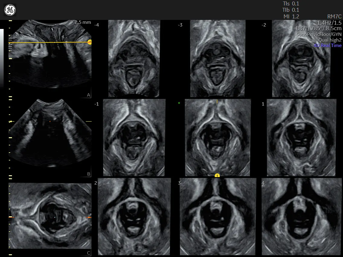
A comprehensive view of the pelvic floor using Voluson Expert 20
SonoPelvicFloor, RM7C probe
In contrast, today's 3D/4D ultrasounds offer a comprehensive view, allowing obstetrician-gynecologists to visualize the complex interactions of pelvic floor muscles, ligaments, and organs. This enhanced visual capability is crucial for a detailed understanding of the pathophysiology of various pelvic floor disorders, leading to more accurate diagnoses and tailored treatment plans.
In his discussion, Dr. Youssef covers the clinical relevance of pelvic floor lesions in particular, highlighting the need for precise diagnosis in managing conditions such as pelvic organ prolapse and urinary incontinence. He provides a detailed walkthrough of how to practically apply 3D/4D ultrasound technology in clinical settings, underscoring the ability of 3D/4D ultrasounds to enable physicians to effectively diagnose both macro- and microtrauma to the pelvic floor muscles.
Dr. Youssef explains the use of volumetric transducers and the role of artificial intelligence (AI) in simplifying the alignment and measurement processes. He illustrates how these advancements enhance diagnostic accuracy and make the examination process more efficient and user-friendly for clinicians.
Exploring the Benefits of New Ultrasound Technology
Dr. Youssef elaborates on how modern 3D/4D ultrasound technologies streamline the traditionally complex pelvic floor examination process. He points out that the complexity of pelvic floor assessments has been a significant challenge, primarily due to the intricate anatomy and the need for precise imaging.
New ultrasound technologies, particularly 3D/4D imaging, simplify this process by providing clearer and more detailed views of the pelvic floor structures. This allows for a more straightforward and less time-consuming examination, reducing the complexity that has long been associated with pelvic floor diagnostics.
Another significant benefit Dr. Youssef discusses is incorporating AI in ultrasound technology. AI-driven workflow guidance and automated alignment have substantially improved the efficiency and accuracy of pelvic floor assessments. This technology aids in ensuring precise plane alignment and measurements—crucial factors for accurate diagnosis. Dr. Youssef notes that AI not only guides clinicians through the examination process but also automates many of the previously manual and time-consuming steps, thereby enhancing the overall efficiency of the diagnostic procedure.
Dr. Youssef cites statistics indicating a significant reduction in exam time using new ultrasound technologies. According to data from Voluson, these advanced methods can reduce examination time by up to 87% compared to manual examinations. This drastic reduction is beneficial for clinicians in terms of efficiency and enhances the patient experience by minimizing the duration of the examination. This improvement in speed, without compromising the quality of the assessment, represents a major step forward in pelvic floor diagnostics.
Introducing the SonoPelvicFloor Tool
Dr. Youssef introduces the SonoPelvicFloor tool as a significant advancement in pelvic floor ultrasound technology, highlighting its integration of AI to simplify the examination process. This innovative tool streamlines pelvic floor assessments by automating plane alignment and measurements—tasks that previously required meticulous manual effort and expertise. Such automation reduces the time taken for each exam and minimizes the potential for human error, enhancing both efficiency and accuracy.
The SonoPelvicFloor tool's ability to provide real-time, automated measurements and alignments is crucial for conducting precise and consistent evaluations. This precision is particularly vital for diagnosing conditions such as levator ani muscle avulsion and assessing the integrity of pelvic floor muscles. By expediting the examination process and improving the quality of diagnostic data, the tool significantly contributes to more informed clinical decisions.
A real-time, 4D view of the pelvic floor using SonoPelvicFloor Voluson Expert 20, RAB6
Furthermore, the SonoPelvicFloor tool is instrumental in reducing uncertainties in pelvic floor anatomy analysis. By offering clear and accurate visualizations, it aids in identifying and evaluating anatomical variations and pathologies, which can be challenging to discern with traditional methods. This feature is invaluable in complex cases, where detailed anatomical information is essential for accurate diagnosis and treatment planning.
In clinical practice, the SonoPelvicFloor tool enhances the confidence of obstetricians-gynecologists in diagnosing and formulating treatment strategies. Its advanced capabilities enable clinicians to navigate the intricacies of pelvic floor anatomy more effectively, marking a significant improvement in patient care. Additionally, AI enhancement could allow more patients to be seen in a day while lowering clinical overhead by reducing the need for senior physician oversight of new trainees and fellows.
Dr. Youssef emphasizes that the tool represents more than just a technological breakthrough; it is a vital development in empowering clinicians to address pelvic floor disorders more effectively and efficiently.
A Significant Leap Forward for Pelvic Floor Ultrasound
The evolution of ultrasound technology, as expertly elucidated by Dr. Youssef, represents a substantial leap forward in pelvic floor diagnostics. This advancement, from basic 2D to sophisticated 3D/4D and AI-integrated imaging, has revolutionized the way obstetricians-gynecologists approach pelvic floor assessments. The SonoPelvicFloor tool, in particular, exemplifies this progress, offering unprecedented accuracy and efficiency in diagnosing and managing complex pelvic floor conditions.
By simplifying examination procedures, reducing exam time, and providing a clearer understanding of pelvic floor anatomy, these technological innovations enhance clinical practices and pave the way for improved patient outcomes in pelvic health.
Dr. Youssef’s lecture can be viewed here: 3D ultrasound of the pelvic floor

