Assessing Uterine Contractility with Ultrasound Before IVF Embryo Transfer
Embryo transfer represents the culmination of the IVF journey, bringing together embryo quality, endometrial receptivity, and the uterine environment at a critical moment in the reproductive process. While clinicians have traditionally focused on embryo quality, endometrial thickness, and the timing of the implantation window, growing evidence suggests that another factor may also influence success: uterine contractility.¹⁻⁷
Once considered simply part of normal reproductive physiology, uterine contractility is increasingly recognised as a measurable characteristic of the uterine environment that may impact embryo implantation and IVF outcomes.¹⁻⁵ Even when embryo quality is high and endometrial thickness appears optimal, elevated myometrial activity has been associated with lower implantation rates.¹˒⁴˒¹⁰
Today, transvaginal ultrasound (TVUS) offers clinicians the ability to visualise and assess uterine activity in real time during the implantation window, complementing established measures of endometrial receptivity.⁵˒¹⁰ As with many advanced ultrasound applications, obtaining meaningful information depends on clinician expertise in image acquisition and interpretation.
Understanding the Physiology of Uterine Contractility
Throughout the menstrual cycle, the uterus undergoes rhythmic, wave-like myometrial peristalsis.⁹˒¹⁰ These contractions are influenced by fluctuations in estrogen and progesterone, as well as by the local effects of oxytocin and prostaglandins.¹⁰
The activity originates within the junctional zone (JZ), the specialised inner layer of the myometrium surrounding the endometrium, and can be visualised using high-resolution transvaginal ultrasound.¹¹ Unlike labour contractions, JZ peristalsis is a continuous physiological process, with contraction frequency and direction changing throughout the menstrual cycle.¹¹
During the late follicular and periovulatory phases, rising estrogen levels promote contractions moving from the cervix toward the fundus, supporting sperm transport.⁹ Following ovulation, progesterone suppresses contraction frequency, creating the relatively quiescent uterine environment thought to facilitate blastocyst implantation.⁴˒¹⁰
Understanding these physiological changes provides important context when evaluating uterine activity before embryo transfer.
Evidence Linking Uterine Contractility and IVF Outcomes
A growing body of research has identified an association between elevated uterine contractility at the time of embryo transfer and lower implantation and clinical pregnancy rates.¹˒⁴ Increased uterine activity may mechanically displace the embryo or alter the endometrial environment during the implantation process.⁷˒⁹˒¹⁰
Initial studies focused primarily on contraction frequency. More recent investigations have expanded the assessment to include wave direction, amplitude, velocity, and the overall coordination of uterine activity.⁴˒⁵˒¹⁰
While the clinical significance of these dynamic characteristics continues to be explored, assessment of uterine contractility may offer additional information to support individualised embryo transfer decisions.⁴˒¹⁰
Clinical Interpretation in Context
As evidence continues to emerge, uterine contractility assessment should be viewed as one component of a comprehensive fertility evaluation. Its value depends not only on the ability to visualise uterine activity, but also on the clinician's expertise in interpreting findings within the broader clinical picture.⁴˒¹⁰
Contractility measurements should be considered alongside embryo quality, endometrial characteristics, patient history, and other relevant clinical factors rather than in isolation.⁴˒¹⁰˒¹¹
Although no universally accepted contraction frequency threshold exists, several studies have reported reduced implantation rates when uterine activity exceeds approximately three contractions per minute immediately before embryo transfer.¹˒⁴ When elevated activity is identified, treatment decisions should be guided by the overall clinical context.
Pharmacologic approaches, including oxytocin antagonists, have been investigated, but evidence supporting routine intervention remains limited, and standardised management strategies have not yet been established.⁴˒¹⁰
Research has also suggested that initiating luteal-phase progesterone support at oocyte retrieval, rather than at embryo transfer, may help reduce contraction frequency by the day of transfer.² In cases where uterine activity remains elevated despite intervention, some clinicians consider postponing transfer to a future frozen embryo transfer cycle, although this approach has not yet been evaluated in dedicated clinical trials.
Assessing Uterine Contractility with Ultrasound
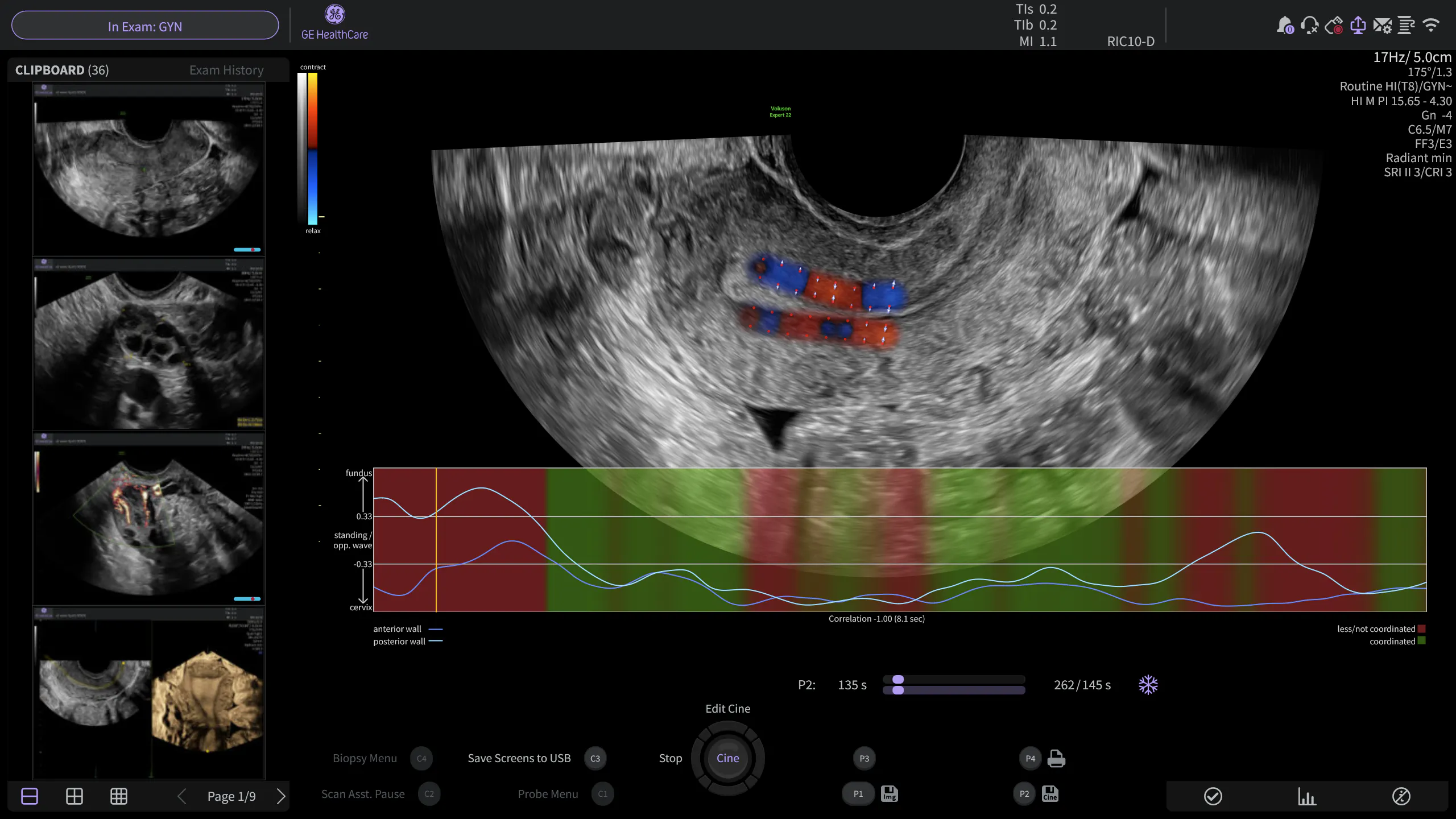
Assessment is typically performed immediately before embryo transfer using high-resolution transvaginal ultrasound and real-time cine imaging.⁵
Many published studies use observation periods of approximately four minutes to evaluate low-frequency subendometrial contractions.⁵˒⁸ During this assessment, clinicians evaluate:
- Contraction frequency
- Wave direction
- Overall coordination of uterine activity
This real-time cine evaluation provides a functional view of uterine physiology that complements traditional pre-transfer ultrasound findings. By capturing dynamic uterine activity, ultrasound can offer information that static imaging alone cannot provide.¹⁰
Bringing Functional Assessment into Clinical Practice
Unlike many conventional ultrasound measurements, uterine contractility can be assessed dynamically in real time.³˒⁸˒¹⁰ Transvaginal ultrasound allows clinicians to visualise and quantify this activity, offering valuable insight into an often-overlooked aspect of the uterine environment.
As technology continues to evolve, quantitative techniques such as speckle-tracking analysis aim to reduce reliance on visual wave counting and provide more objective assessments. However, additional multicentre validation is still required before widespread clinical implementation.
Regardless of future technological advances, clinician expertise will remain central to successful assessment. High-quality image acquisition, thoughtful interpretation, and integration of findings with other clinical factors are essential to ensuring that uterine contractility assessment contributes meaningfully to patient care and informed clinical decision-making.
Expanding Understanding of the Uterine Environment
The ability to visualise uterine activity adds a new dimension to pre-transfer assessment, helping clinicians better understand the complex physiological environment in which implantation occurs. While research continues to define the role of uterine contractility in IVF outcomes, transvaginal ultrasound provides a non-invasive way to evaluate this dynamic process in real time.
As knowledge advances, incorporating functional assessment alongside traditional measures of embryo and endometrial evaluation may further support personalised fertility care and optimise decision-making throughout the IVF journey.
References:
1.Fanchin R, Righini C, Olivennes F, de Ziegler D, et al. Uterine contractions at the time of embryo transfer alter pregnancy rates after in-vitro fertilization. Hum Reprod. 1998;13(7):1968-1974. doi:10.1093/humrep/13.7.1968
2.Fanchin R, Righini C, de Ziegler D, Olivennes F, Ledée N, Frydman R. Effects of vaginal progesterone administration on uterine contractility at the time of embryo transfer. Fertil Steril. 2001;75(6):1136-1140. doi:10.1016/s0015-0282(01)01787-3
3.Zizolfi B, Rees CO, Musone M, et al. Quantitative ultrasound measurement of uterine contractility in septate uterus vs normal uteri: a multicenter prospective study. Fertil Steril. 2025;124(4):728-736. doi:10.1016/j.fertnstert.2025.05.159
4.Vidal A, Trejos V, von Wolff M, et al. Lower pregnancy rate in women with high uterine peristalsis before embryo transfer: a systematic review and meta-analysis. Reprod Biol Endocrinol. 2025;23(1):49. doi:10.1186/s12958-025-01380-5
5.Rees CO, De Boer A, Huang Y, et al. Uterine contractile activity in healthy women throughout the menstrual cycle measured using a novel quantitative two-dimensional transvaginal ultrasound speckle tracking method. Reprod Biomed Online. 2023;46(1):115-122. doi:10.1016/j.rbmo.2022.08.104
6.Liu KE, Hartman M, Hartman A, Luo ZC, Mahutte N. The impact of a thin endometrial lining on fresh and frozen-thaw IVF outcomes: an analysis of over 40 000 embryo transfers. Hum Reprod. 2018;33(10):1883-1888. doi:10.1093/humrep/dey281
7.Craciunas L, Gallos I, Chu J, et al. Conventional and modern markers of endometrial receptivity: a systematic review and meta-analysis. Hum Reprod Update. 2019;25(2):202-223. doi:10.1093/humupd/dmy044
8.Rees CO, Thomas S, de Boer A, et al. Quantitative ultrasound measurement of uterine contractility in adenomyotic vs normal uteri: a multicenter prospective study. Fertil Steril. 2024;121(5):864-872. doi:10.1016/j.fertnstert.2024.01.009
9.Kunz G, Beil D, Deininger H, Wildt L, Leyendecker G. The dynamics of rapid sperm transport through the female genital tract: evidence from vaginal sonography of uterine peristalsis and hysterosalpingoscintigraphy. Hum Reprod. 1996;11(3):627-632. doi:10.1093/humrep/11.3.627
10.Nicolì P, Viganò P, de Ziegler D, et al. Decoding uterine contractility: from physiology to pathology, through emerging technologies. Reprod Biomed Online. 2026;52(3):105355. doi:10.1016/j.rbmo.2025.105355
11.Kuijsters NP, Methorst WG, Kortenhorst MS, Rabotti C, Mischi M, Schoot BC. Uterine peristalsis and fertility: current knowledge and future perspectives: a review and meta-analysis. Reprod Biomed Online. 2017;35(1):50-71. doi:10.1016/j.rbmo.2017.03.019

