Benefits of Ultrasound with the SonoL&D Tool
Clinicians understand the benefits of ultrasound during pregnancy, but technological advances have expanded many of ultrasound's advantages into the delivery room. Labor is a stressful time, for the patient and clinicians. It can move painfully slowly or happen in a blink. Portable ultrasound monitors with smaller footprints are easier than ever to integrate into labor and delivery to monitor the patient's progress. They offer valuable information to clinicians and patients, as well as automated measurements to save you time conducting exams.
Ultrasound provides vital information throughout pregnancy and labor, including fetal position, placenta location, amniotic fluid evaluation, and fetal cardiac activity. During active labor, ultrasound can support decision-making with objective measurements that track progression over time. Digital exams to determine fetal progression and positioning are helpful but can be imprecise and increase the risk of infection. Ultrasound can precisely visualize fetal progression and position, among other measurements, to monitor the patient's progress and make informed clinical decisions about delivery.
The VolusonTM SonoL&D tool provides objective assessments during labor in an easy-to-use machine. The SonoL&D tool complements manual exams to provide more details on:
- Fetal presentation
- Labor progress
- Cervical dilatation
- Fetal wellbeing
Benefits of Ultrasound During Labor
Ultrasound exams and the new SonoL&D tool provide you with objective clinical information and visuals, keeping everyone on the same page throughout labor. Tracking progress allows you to identify obstructed labor, overcome uncertainty in clinical exams, and determine if the head is appropriately engaged. The visuals allow you to share progress with the patient and make decisions about surgery or instrumental delivery.
Abdominal palpation does not always identify malpresentation, which is where ultrasound scanning can play a complementary role to clinical assessment. With transabdominal imaging, you can assess the placenta location, fetal presentation, and fetal anatomy. Transperineal imaging tracks fetal progression during the second stage of labor. You can also view fetal head flexion with imaging.
SonoL&D Angle of Progression
SonoL&D Midline Angle Measurement
Ultrasound, with new AI-enabled features and automated measurements, aids in making quick and effective decisions during labor. You have comprehensive, objective information in addition to clinical examination to support next steps, particularly in cases of prolonged labor.
How the SonoL&D Tool Works
The SonoL&D tool is an easy-to-use feature of Voluson ultrasound products. It provides clinical and patient support during the second stage of labor with objective clinical measurements and graphic support to share with patients and partners. The large touchscreen and small footprint of Voluson makes it easy to maneuver in the delivery room and to display informational graphics to keep patients apprised of their progress.
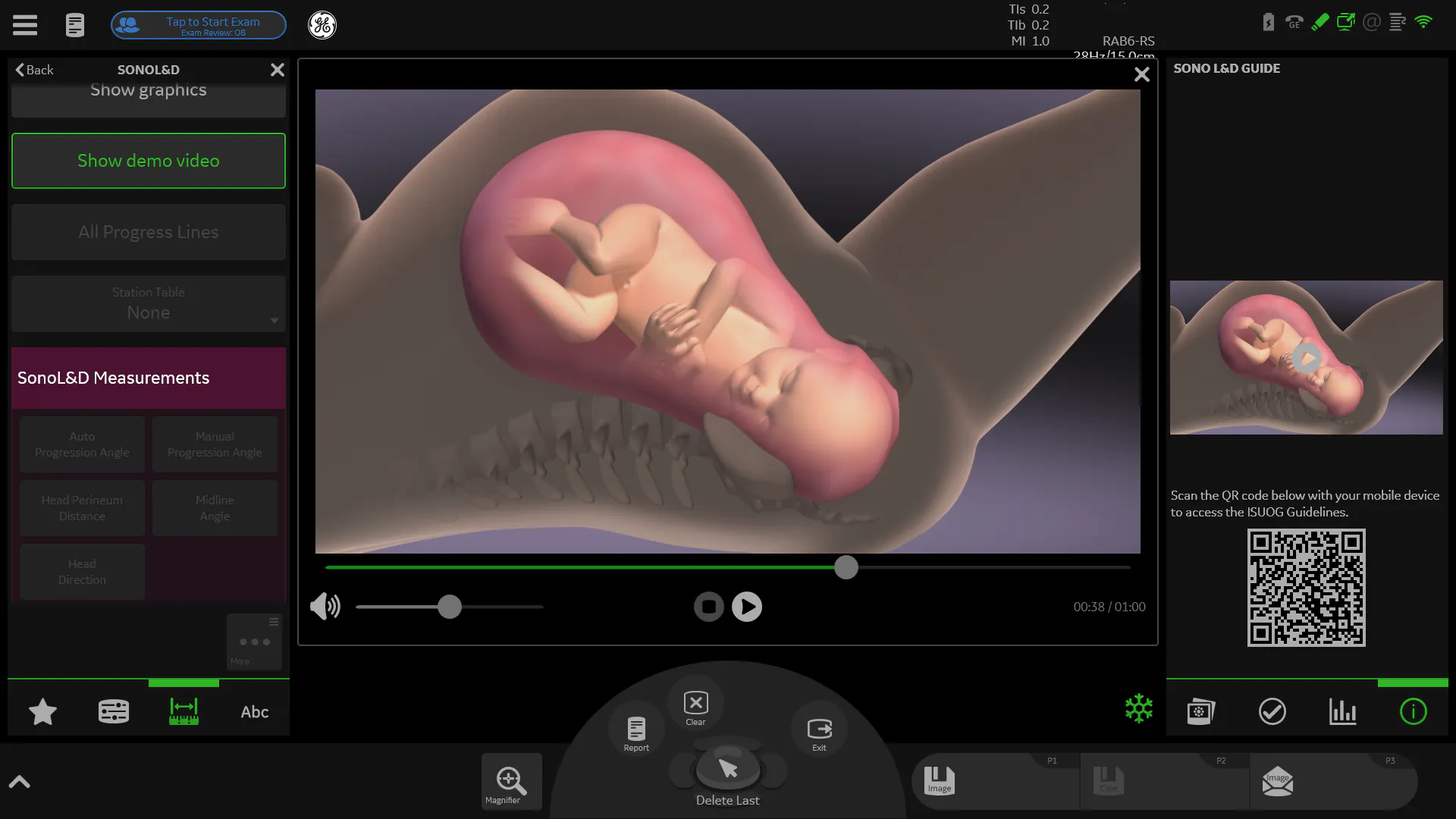
To start, select the box for SonoL&D on the new patient screen. You can come back to the tool and add additional measurements as labor progresses. You can select an educational video on the touch monitor to share with patients. Patients can view a 3D graphic that clearly shows the baby's position and progression.
You can perform two views for the exam: the angle of progression in the sagittal view and the head/perineum distance in the transverse view.
- To measure angle of progression, place the transducer in the mid-sagittal plane on the perineum. When you have the head and pubic bone visualized, freeze the image and select "Calc." You have the option to measure manually or use the automated features in the tool. The automated features help find the proximal and distal ends of the pubic bone, as well as the fetal skull. You can adjust these locations as needed. Once identified, the SonoL&D tool calculates the angle and saves it with a timestamp. The tool also produces a graphic that displays the head progression visually to share with the patient and partner.
- To measure the head/perineum distance, place the transducer on the perineum in a transverse orientation. Measure from the top of the image area to the fetal skull. The tool saves the measurement with a timestamp.
As labor progresses, you can view progress lines based on previous measurements. This data is also added to an L&D worksheet that includes your objective measurements as well as data from clinical assessments, such as station and cervical dilatation. The patient archive within the tool lets you reopen exams, allowing you to move between multiple patients. The SonoL&D tool for the Voluson ultrasound provides features to make scans simpler and user-friendly, while providing quality imaging that lets you care for patients with confidence.

