Best Practices for Viewing and Classifying Congenital Uterine Anomalies
In the field of fertility medicine, there are a number of challenges and controversies surrounding the classification of uterine anomalies. Differentiating between uteri that are normal, septate and T-shaped will be the focus of this article, with other these anomalies not covered. This article aims to distill complex information into a simpler presentation, exploring the nuances of the debate and highlighting relevant and practical points.
Understanding Uterine Classification
Septate and T-shaped uteri can have negative impacts, though still relatively unexplored, in fertility medicine. Despite the noted impacts in pregnancy in the literature associated with congenital uterine anomalies, there is still significant controversy because the definitions of each anomaly are debated.
Thus far, there are conflicting perspectives from various societies, such as the American Society for Reproductive Medicine (ASRM) and European Society of Human Reproduction and Embryology (ESHRE)/European Society for Gynaecological Endoscopy (ESGE). This controversy, and perceived deficiencies with both models, led to the development of the Congenital Uterine Malformation by Experts (CUME) statement published in 2018 and more recent work done by this group.
Assessment of Uterine Anomalies
A structured approach to assessing the uterus should always be a priority. Standard sagittal and transverse views of the uterus form the foundation of our understanding of the uterus and whether it is normal or abnormal. However, traditional approaches that only involve a single sagittal and single transverse image are of the past. Of course, if a sonographer or sonologist identified an abnormality (e.g. a fibroid), that would also be added to the basic required image captures. Nowadays, video captures are essential and, in many centers, are considered to be mandatory, including a sagittal sweep from side to side and a transverse sweep from inferior to superior (or vice versa). Generally, this will mean a sweep from cornua to cornua and cervix to fundus. However, this may not be the case in some congenital abnormalities like unicornuate uteri, seeing as they only have one cornua. In the context of septate or T-shaped uteri, the cornua-cornua and cervix-fundus sweeps are relevant and necessary.
In addition, it is customary for the evaluation of uterine anomalies to use three-dimensional captures, including 3D volume contrast imaging (VCI), to reconstruct a coronal view of the uterus. As you will note below, it is this rendered 3D image output that is essential to characterizing specific features that will be necessary to diagnose (or not) septate or T-shaped uteri.
Septate Uterus Classification
The 2016 ASRM guidelines for identifying septate uteri were based on indentation depth (more than 1.5 cm) and angle (< 90°). Seeing as a normal/arcuate uterus was defined by an indentation depth of < 1 cm and angle > 90°, there was a grey zone of uteri with indentation depths between 1 and 1.5 cm. In 2021, the ASRM updated their congenital anomalies classification to change the indentation depth cut-off to 1 cm, leaving the angle cut-off unchanged. The ESHRE/ESGE publication from 2013 defined a septate uterus as one having an abnormal ratio, whereby the indentation is > 50% of the uterine wall thickness. However, they did not define uterine wall thickness until an updated publication in 2016, and even then, it remained controversial. When comparing the 2016 ASRM guideline with the 2013/2016 ESHRE/ESGE guidelines, there were either patients in a grey zone or a significant overclassification of septate uteri, respectively.
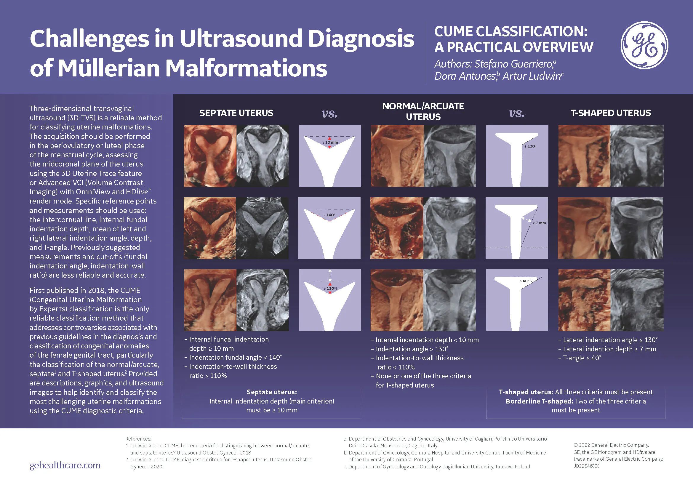
The CUME classification from 2018 utilizes the following criteria: fundal indentation depth ≥ 1 cm, which matches the 2021 ASRM guideline, indentation fundal angle < 140° and indentation-to-wall thickness ratio > 110%. According to the publication, which involved critical assessment of these criteria—as reviewed by clinicians, sonologists, and surgeons—there was very high diagnostic accuracy and agreement between experts. The criteria also yielded what is perceived to be a more reasonable diagnostic prevalence of septate uteri, versus the high (and potentially incorrect) diagnostic rate when using the ESHRE/ESGE classification.
T-Shaped Uterus Classification
Like the septate uterus controversy, recent dialogues within the medical community have revealed significant challenges in accurately classifying T-shaped uteri. Indeed, T-shaped uteri may be even more enigmatic to the medical community, as well as lay community. The absence of standardized and objective diagnostic criteria has led to variations in interpretations among experts, prompting a critical reassessment of existing approaches.
The ASRM 2016 nor 2021 classification systems make reference to T-shaped uteri. However, the ESHRE/ESGE 2013 classification does; it's characterized by a narrow uterine cavity due to thickened lateral walls with a correlation 2/3 uterine corpus and 1/3 cervix. The CUME classification attempted to define a T-shaped uterus in a similar fashion to their study on septate uteri.
From this, researchers have introduced innovative measurements to address the diagnostic challenges associated with T-shaped uteri. These include lateral indentation depth (≥ 7 mm), lateral indentation angle (≤ 130°), and a newly proposed metric known as the T-angle (≤ 40°). Whilst these measurements aim for objectivity, there is only fair to moderate agreement thus far. To overcome the possible challenges with agreement on a single feature, the authors proposed that if patients have all three features, there is a high positive predictive value of truly having a T-shaped uterus.
Research Landscape
We know from a systematic review by Coelho Neto and colleagues that 20 studies spanning from 1979 to the present have been published, more in recent years, encompassing diverse perspectives on prevalence, associations with reproductive outcomes and the impact of surgical interventions. However, in the absence of standardized diagnostic approaches (e.g. three-dimensional ultrasound in all people), standardized and objective diagnostic criteria (e.g. those proposed above by CUME) and interventional studies (e.g. randomized controlled trials) on the basis of the accurate diagnoses, we will have no idea as to the true utility of various outcomes. In fact, because of the dearth of evidence, it is possible that T-shaped uteri should be considered a normal variant, and no interventions should be performed unless they are done so under the lens of a research study.
Enhancing Diagnostic Technologies
Insights into future possible objective measurements, aided by technology—such as HysteroSonoAVC or septation volumetric indices—can also be considered as potentially more accurate and reliable.
Looking Ahead
This article concludes with a call for widely accepted criteria for T-shaped uterine anomalies, reflecting the sentiments expressed in contemporary research. Whilst there may be more worldwide unification around septate uteri, more work still needs to be done. Ongoing discussions and research underscore the need for consensus to guide clinical practices and research endeavors. The complexities surrounding septate and T-shaped uteri emphasize the importance of cautious approaches to interventions until a standardized framework is established.

