

More than 1.3 million babies worldwide are born with a congenital heart defect each year. And you play a critical role in ensuring these children have the best chance at life. When the decisions you make mean the world to a mother and her child, are you willing to settle when it comes to ultrasound technology?
Diagnosing Congenital Heart Defects with Ultrasound Technology
A Clearer Picture of Fetal Heart Abnormalities
Congenital heart defects are difficult to detect. GE Healthcare has created revolutionary technology for assessing and monitoring the fetal heart.

More than 1.3 million babies worldwide are born with a congenital heart defect each year. And you play a critical role in ensuring these children have the best chance at life. When the decisions you make mean the world to a mother and her child, are you willing to settle when it comes to ultrasound technology?
No one wants to hear that their baby has a heart defect. And no one wants to deliver that distressing news. Barbara Del Prince vividly remembers those gripping moments with her patients when she was performing ultrasounds as a sonographer in the late 80s.
“Nothing is more devastating than when you’re evaluating a patient who expects to have a completely normal pregnancy, and you see something that could be potentially life-threatening to the fetus. It’s a very emotional time for the patient, the partner, and the physician taking care of her,” Del Prince stressed.
Congenital heart defects are the most common congenital defects, affecting 1 out of every 110 babies born worldwide.1 Still, most women are completely blindsided by the diagnosis because 90% occur in pregnancies where there are no known risk factors.2
Congenital heart defects are also among the most difficult fetal abnormalities to detect. In general, low-risk detection rates are between 30 to 50 percent.³ GE Healthcare is committed to changing those numbers with its Voluson™ technology.
“That’s one reason why we are so focused on the fetal heart with Voluson,” said Del Prince, who is Clinical & Marketing Director for GE Healthcare, Women’s Health Ultrasound
“We want to make assessment easier and more accessible for every physician, to ultimately bring undetected rates down to as close to zero as we can.”
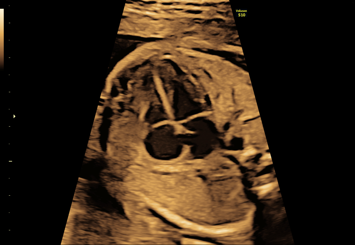
26 week 4 chamber view of fetal heart
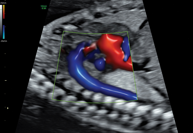
Voluson Radiantflow with HDRes aortic arch
GE Healthcare has created revolutionary technology for assessment, diagnosis, and monitoring the fetal heart. With ultra-fast volume rates, flexible imaging formats and excellent resolution of electronic 4D to the evaluation of fetal heart structure using SonoVCAD heart, Voluson offers progressive tools that can help physicians provide confident patient answers in less time. Using Radiantflow, physicians can quickly assess even the tiniest vessels, setting a new standard of color Doppler. Physicians can see structural abnormalities as early as 8 weeks, when the fetus is barely the size of a kidney bean.
Del Prince says GE Healthcare continues to push the envelope developing technology that will change the way physicians evaluate the fetal heart. The new Voluson tools are designed to provide valuable clinical information that will give physicians a better indication of the true health of the baby.
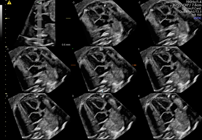
SonoVCAD™heart provides access to essential views of the fetal heart from a single STIC volume.
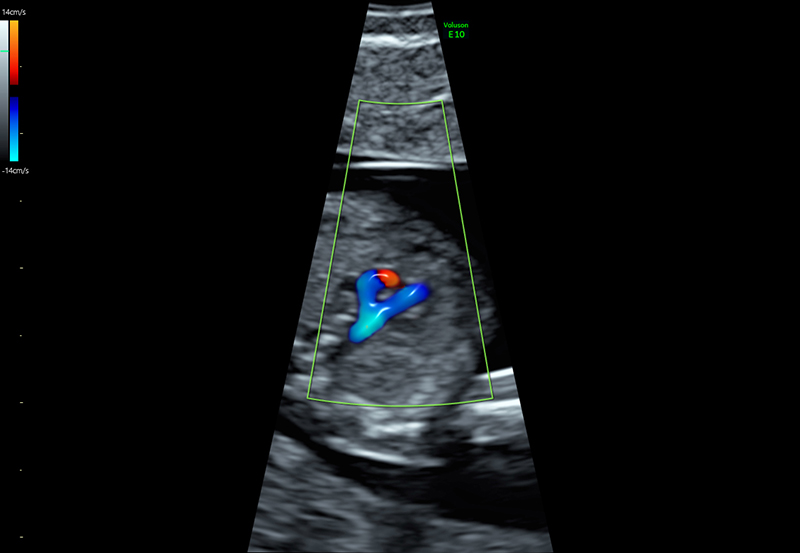
3 Vessel View in a 12 week fetus using Radiantflow
While nothing can lessen that blow of a difficult diagnosis, Voluson technology provides physicians with the knowledge and insight to help patients who are faced with tough choices.
“If there is a fetal heart abnormality—being able to identify it allows patients to make more informed decisions. These can be critical life and death decisions.” Del Prince added, “Physicians can also create a better treatment plan, and be more prepared for delivery.”
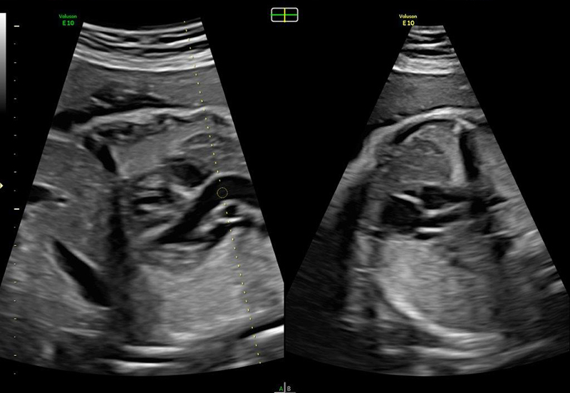
Aorta in the fetal heart seen in 2 planes simultaneously with BiPlane - advanced electronic 4D technology
Voluson HD-Flow™ aortic arch
-
Learn more about how Voluson Sono-Automation technology can help simplify the fetal heart assessment and help provide answers, faster.
-
With Voluson e4D technology, ultrasound image quality is enhanced dramatically by almost completely eliminating artifacts. ‘Imaging of the fetal heart has been revolutionized’ (Dr. Greggory DeVore, 2017)
-
Explore Voluson 3D Printing for clinical prototyping, research and enhanced patient communication – full mesh exports directly from your Voluson system.

