

Diagnosing fetal heart abnormalities can improve the chances of survival for newborns with certain types of congenital heart defects.¹ ² ³ ⁴ But these outcomes are dependent upon your ability to get the most comprehensive evaluation of the fetal heart—to see more, sooner.
Using Ultrasound to Detect Fetal Cardiac Anomalies
Diagnosing Fetal Heart Abnormalities in the First Trimester
A Q&A with Dr. Rabih Chaoui of the Center for Prenatal Diagnosis and Human Genetics on using ultrasound to detect fetal cardiac anomalies.

Diagnosing fetal heart abnormalities can improve the chances of survival for newborns with certain types of congenital heart defects.¹ ² ³ ⁴ But these outcomes are dependent upon your ability to get the most comprehensive evaluation of the fetal heart—to see more, sooner.
Dr. Rabih Chaoui of the Center for Prenatal Diagnosis and Human Genetics in Berlin talks about the power and precision of Voluson™ in diagnosing fetal heart defects as early as 12 weeks.
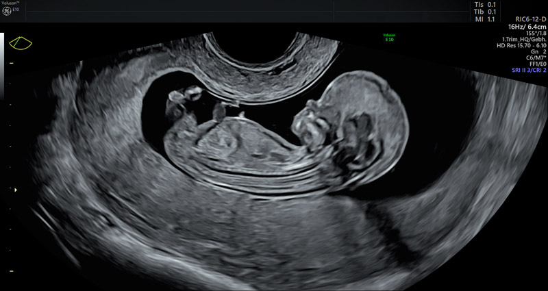
Q: What does Voluson technology allow you to see in fetuses as early as 12 weeks and how does that help in the diagnosis of certain heart defects?A: Examining the fetal heart around 12 weeks of gestation should include the demonstration of the upper abdomen with the situs, the 4-chamber view both in gray scale and color, and the great vessels in the 3-vessel-trachea view in color Doppler. With these simple planes, the majority of complex cardiac anomalies can be ruled out at this stage of gestation.
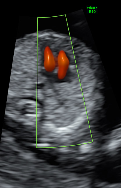
Easy, fast visualization of the tiniest vessels with Voluson color Doppler with Radiantflow.
Along with user experience, technology is a key factor in examination. Voluson´s transabdominal and transvaginal transducers are high-resolution probes, which allow for a very precise diagnosis by the combination of gray scale, color Doppler, and 3D ultrasound.
Q: How did the introduction of Voluson’s Radiantflow change the way you evaluate the fetal heart in early pregnancy? What are the benefits of this tool?A: The advent of Radiantflow has contributed to an increase in the sensitivity of color Doppler—leading to a more accurate diagnosis. The main advantages are a more reliable and precise visualization of flow across the cardiac chambers and great vessels. Increased sensitivity also allows for the demonstration of tiny vessels, including pulmonary veins or the subclavian arteries. With Radiantflow, I’m able to reach a clinical decision faster, reducing examination time.
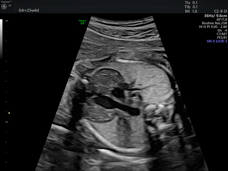
26 week fetal thymus gland.
Q: Why are these ultrasound scans so important in the monitoring and treatment of congenital heart defects?A: Diagnosing cardiac anomalies in early gestation enables patients to get counseling, complete additional testing, and also provides more time for decision making. In most cases, an early diagnosis allows patients to search and contact a clinic with optimal facilities for neonatal cardiac intervention and cardiac surgery. All these steps depend on an accurate diagnosis, which is based on precise information provided by the ultrasound equipment.
Q: How has Voluson technology impacted your practice?A: I have been using the Voluson technology for nearly 15 years and the ongoing improvement of the hardware and software including 3D-technology, color Doppler, and enhanced gray-scale information has allowed me to get more precise diagnoses, even in early gestation.
3 week fetal profile.
With the detailed images, I’ve had the opportunity to observe new anatomical structures including the thymus, spleen, and brain. The high-resolution probes have made it possible to see abnormal course of the subclavian artery, which is reflected in my published research. The technology has also allowed us to discover that intracranial translucency is an early sign of spina bifida and that the maxillary gap indicates facial clefts. And with the continuous stream of innovation in transvaginal probes, we are pushing to make important diagnoses in fetal medicine even earlier than 11 weeks of gestation.
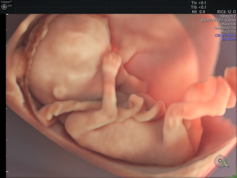
Fetus rendered with Voluson HDlive Studio.
Q: Many physicians are committed to delivering a better patient experience? What role does ultrasound play in providing more value to patients?A: Ultrasound advancements allow us to act as a translator of the world of the womb, which provides a meaningful experience for our patients. In the past, 2D images of abnormalities required a great deal of explanation, but high-resolution 3D/4D technology helps patients really see and better understand the diagnosis. It builds trust, which leads to a better connection with our patients.
-
Learn more about how Voluson Sono-Automation technology can help simplify the fetal heart assessment and help provide answers, faster.
-
Explore here how the Voluson E10’s innovative technologies can help you focus on early prevention.
-
Download a free poster detailing some of the most important facts about fetal heart abnormalities for your examination room or office.
-
Explore here how Voluson 3D Printing for clinical prototyping can support your research efforts and enhance communication with your patient around fetal heart abnormalities.

