

There are more than 35 congenital heart defects—ranging from simple conditions without symptoms, to complex cases that could pose severe, life-threatening problems. You know the challenges of diagnosis, but what about the possibilities of improving detection with electronic 4D technology?
Uncovering Fetal Heart Defects with Ultrasound
Uncovering Fetal Heart Defects with Electronic 4D Technology
Learn about the possibilities of uncovering fetal heart defects and improving detection with electronic 4D ultrasound technology.

There are more than 35 congenital heart defects—ranging from simple conditions without symptoms, to complex cases that could pose severe, life-threatening problems. You know the challenges of diagnosis, but what about the possibilities of improving detection with electronic 4D technology?
Electronic 4D probe technology is not a new concept for maternal fetal medicine specialist Kai-Sven Heling, M.D. He’s been scanning patients with the probe for almost daily for several years—yet he’s still impressed when he sees the images.
“I’m always surprised by the resolution. It’s so clear and you can get perfect color. With the electronic probe you have much better resolution than 2D compared with all the mechanical probes,” said Dr. Heling, from the Prenatal Diagnostics and Therapy in Berlin.
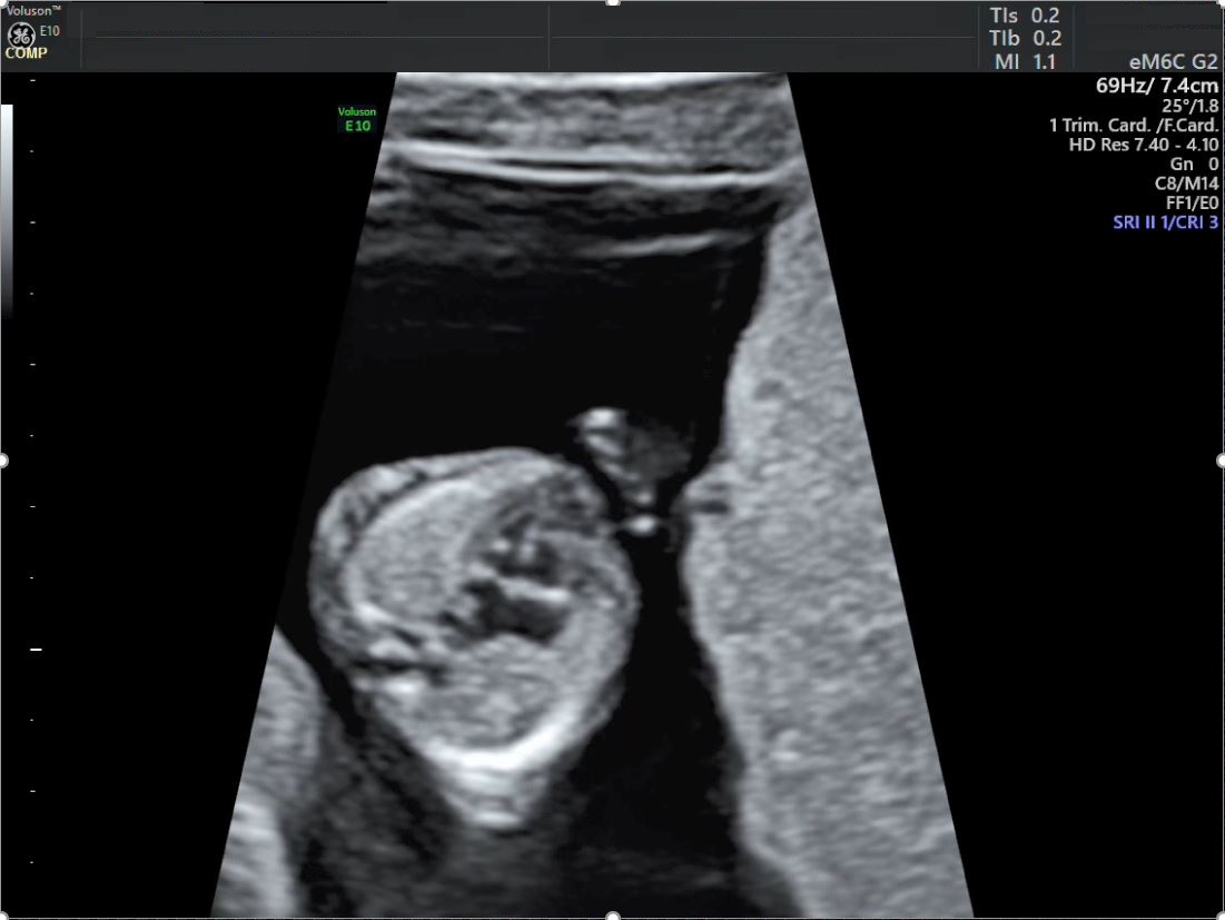
B-Mode image of 4 chamber view in 14 weeks of pregnancy (image courtesy of Dr. Kai Sven Heling)
Heling is referring to the eM6C probe, the world’s first commercially available curved electronic 4D probe from GE Healthcare. With volume rates up to 29 times higher than mechanical probes, the eM6C probe produces real-time display of motion—making it easier to visualize anatomical structures and functionality for a more comprehensive fetal echocardiography.
“It’s changed the way I scan. I do all of my scans with the electronic probe—every patient. For me, the resolution and the images are so much better now than with a mechanical probe. And of course, the Bi-Plane feature only works with the electronic probe,” Heling said.
Bi-Plane imaging allows for the simultaneous display of images in two perpendicular planes. With the Voluson™ E10 ultrasound, Bi-Plane imaging is just one of the many unique tools that can help you achieve valuable information, faster. Not only can you see the heart in greater detail, but it takes less time to perform measurements, and the format can increase clinical confidence by serving as a guide.
“In the past, it was sometimes difficult to find structures like the aortic arch. With Bi-Plane, you have one image where the camera line is in the vessel—and then the second image you have the correct arch—which means you are completely 100% clear that you are at the correct place,” Heling explained.
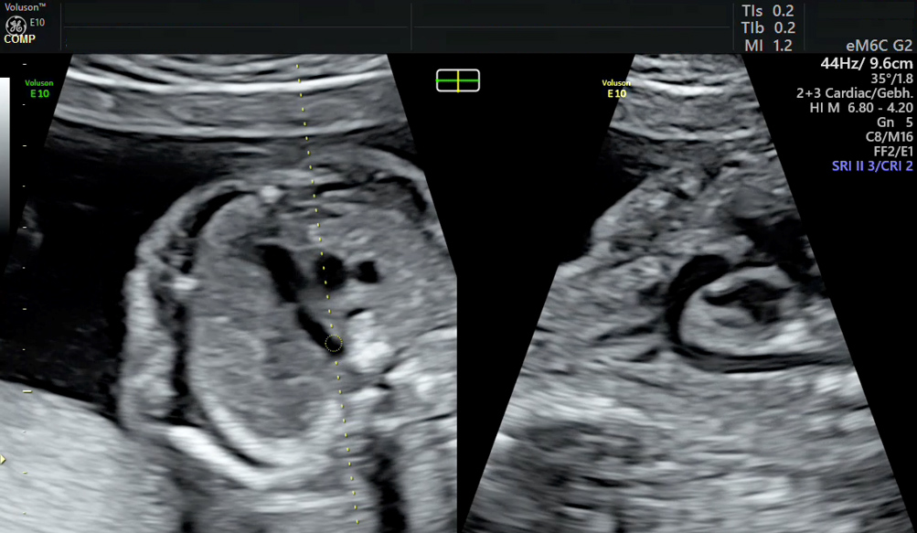
Bi-Plane image showing the aortic arch in B-Plane (image courtesy of Dr. Kai Sven Heling)
The mode also makes it easier to view the ventricular septum, and potential anomalies in the wall between the main pumping chambers. Ventricular Septal Defect, or VSD, is the most common congenital cardiac defect in children, and accounts for 37 percent of cases.1,2,3 During an assessment where Bi-Plane is used, physicians may see what they suspect to be a hole in the fetus’s heart. If they then take the added step of switching on color Doppler, it allows them to evaluate blood flow which helps confirm a VSD diagnosis.
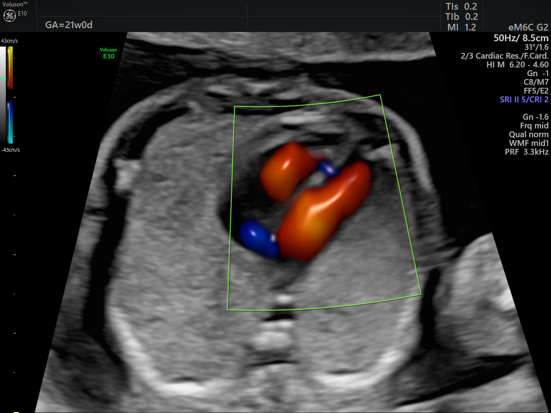
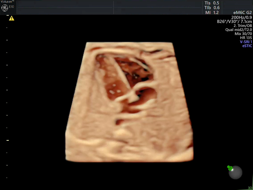
Ventricular Septum Defect (VSD) with Radiantflow™
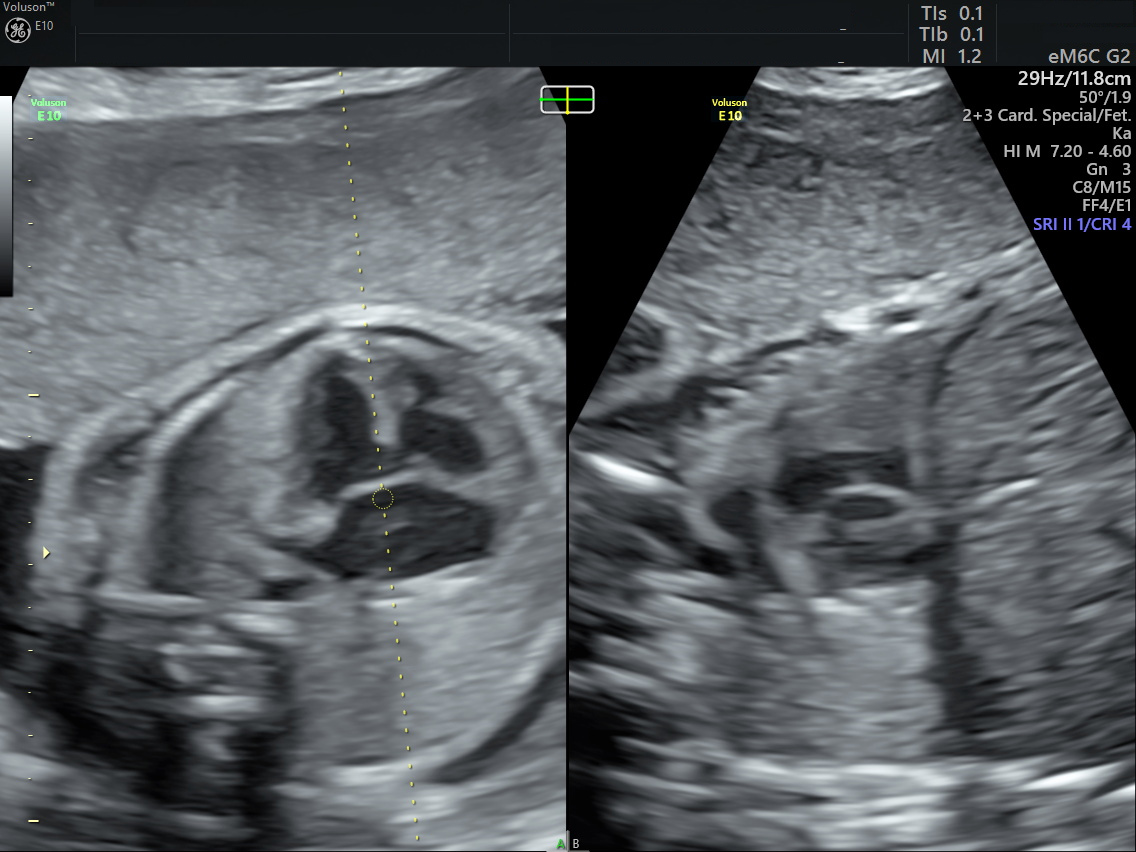
Ventricular Septum in Artrio Ventricular Septum Defect displayed in Bi-Plane Mode (image courtesy of Dr. Kai Sven Heling)
You can also enhance your clinical capabilities in Bi-Plane mode by using Voluson’s Volume Contrast Imaging. VCI-A delivers extraordinary contrast resolution through thick slice volume of grey scale and color Doppler images. It can be a huge advantage in fetal heart assessment—especially if the baby isn’t in the ideal position for scanning.
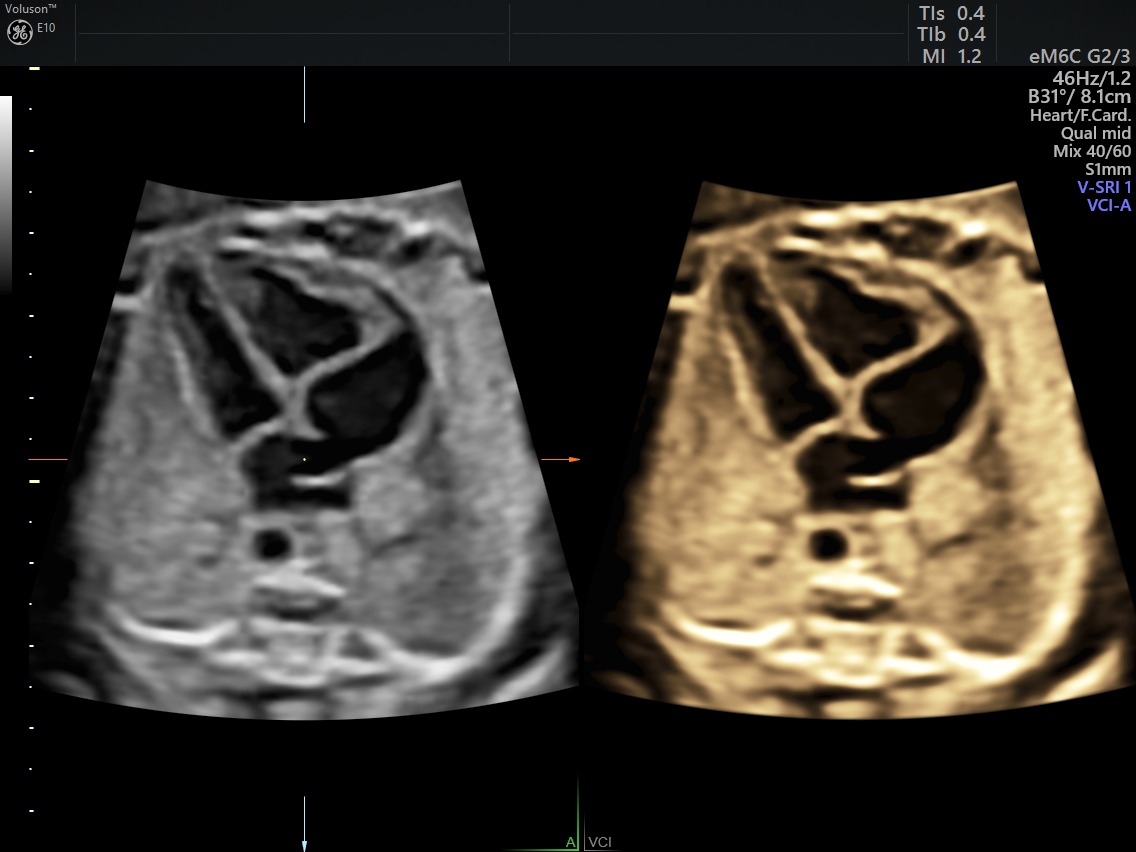
4 chamber view with VCI-A
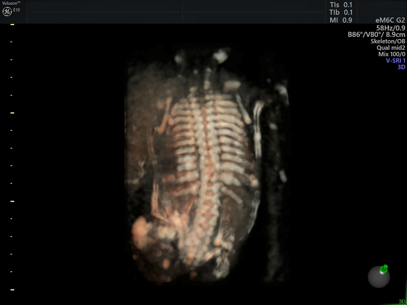
Visualization of the spine with HDlive Silhoutte (image courtesy of Dr.Kai Sven Heling)
“If the baby is on its belly and shows us its spine—it’s more difficult to make a diagnosis. In such cases, you can use Volume Contrast Imaging, because the resolution is so much better. You can clearly demonstrate differentiation between anatomy much better than in an original 2D scan,” Heling said.
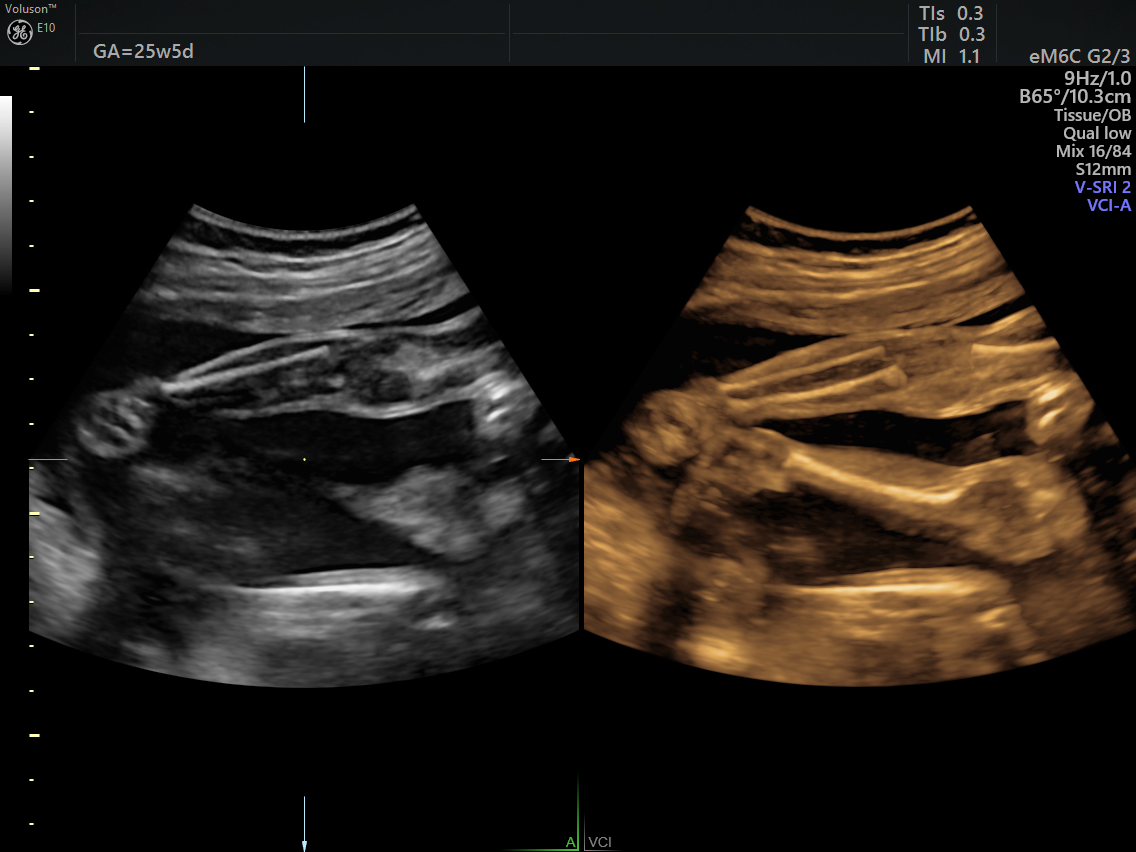
Lower extremities, clearly showing the advantages of Volume Contrast Imaging
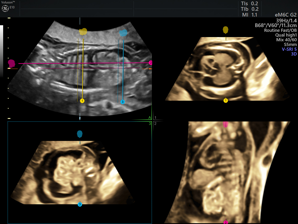
VCI-OmniView, displaying three different lines through the thorax, and the abdomen
Dr. Heling is also quick to point out these features are available with the touch of a button. The e4D technology was also designed as a path to better efficiency—and the tools can have a significant impact , not only on your ability to see anatomy clearly, but also on your workflow.
“It’s very fast. I can scan and store hundreds of images for one exam. With the Voluson ultrasound systems, the workflow is perfect.” Heling concluded, “I always want to create good images of the fetus. A good image of the face, brain and heart are important to me and to the parents. Exclusive e4D technologies on my Voluson system, like Bi-Plane, eSTIC and SnapShot allow me to see important fetal anatomy, and I’m happy about that.”
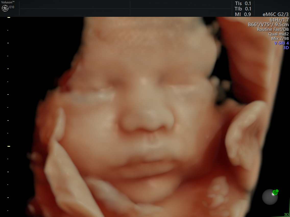
Surface Rendering of the face
4 chamber view with HDlive Rendering (images curtesy of Dr. Kai Sven Heling)
-
Download to learn more about Voluson™ electronic 4D innovations.
Ghosh S, Sridhar A, Solomon N, Sivaprakasham M. Transcatheter closure of ventricular septal defect in aortic valve prolapse and aortic regurgitation. Indian Heart J. 2018 Jul - Aug;70(4):528-532.
Hopkins MK, Goldstein SA, Ward CC, Kuller JA. Evaluation and Management of Maternal Congenital Heart Disease: A Review. Obstet Gynecol Surv. 2018 Feb;73(2):116-124.
Kenny D. Interventional Cardiology for Congenital Heart Disease. Korean Circ J. 2018 May;48(5):350-364.

