

The fetal heart is complicated—but the ultrasound technology that evaluates it, is getting easier.
With half or more congenital heart defects missed prenatally each year¹, do you have the right tools to help save your most precious patients?
New Technology Could Help Detect Critical Fetal Heart Defects Sooner
The Power of Sonography
Learn how GE is creating technology that eliminates some of the guesswork when it comes to the initial assessment of the fetal heart.

The fetal heart is complicated—but the ultrasound technology that evaluates it, is getting easier.
With half or more congenital heart defects missed prenatally each year¹, do you have the right tools to help save your most precious patients?
At 18 weeks, a baby’s heart is the size of an olive and beating about 150 times per minute. The structure itself is extremely complex—and with the baby in constant motion, it’s always a moving target.
Evaluating the fetal heart is complicated. Maternal fetal medicine specialist, Dr. Alfred Abuhamad, says the initial assessment is one of the most difficult ultrasound exams to perform—especially for less-experienced users with minimal training. He’s spent years working with GE to create breakthrough technology that takes some of the guesswork out of the scan.
“We are probably picking up 50 to 60 percent of congenital heart disease prenatally in the United States,” Abuhamad said. “If we want to improve detection before delivery, we really have to make the ultrasound simpler for the people who are doing the basic obstetric ultrasound assessment.”
The solution: Voluson’s™ SonoVCAD™heart.* The newly enhanced tool was developed to assist the non-expert user by acquiring a volume of data and locating and displaying the recommended planes. This takes the user variability out of the equation.
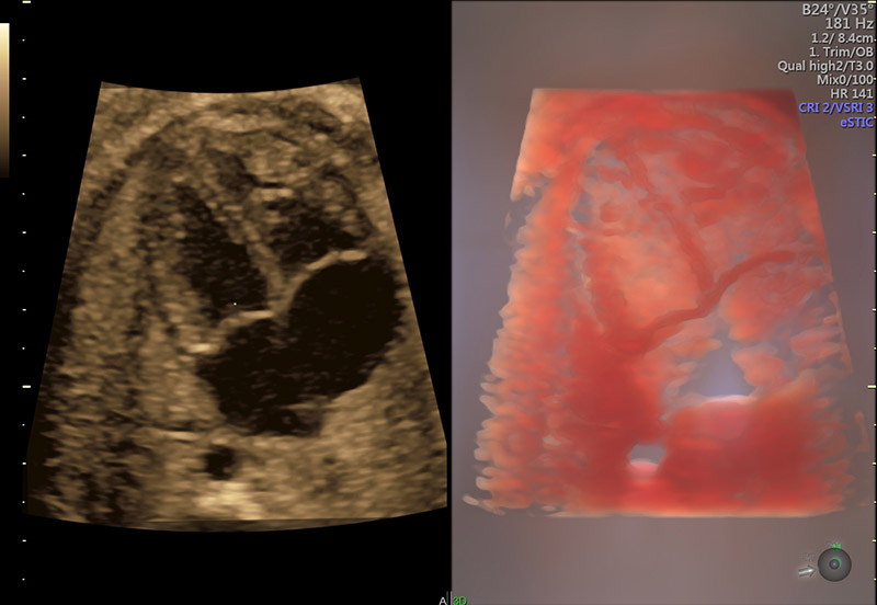
eSTIC with internal structures illuminated with Voluson HDlive™ Studio
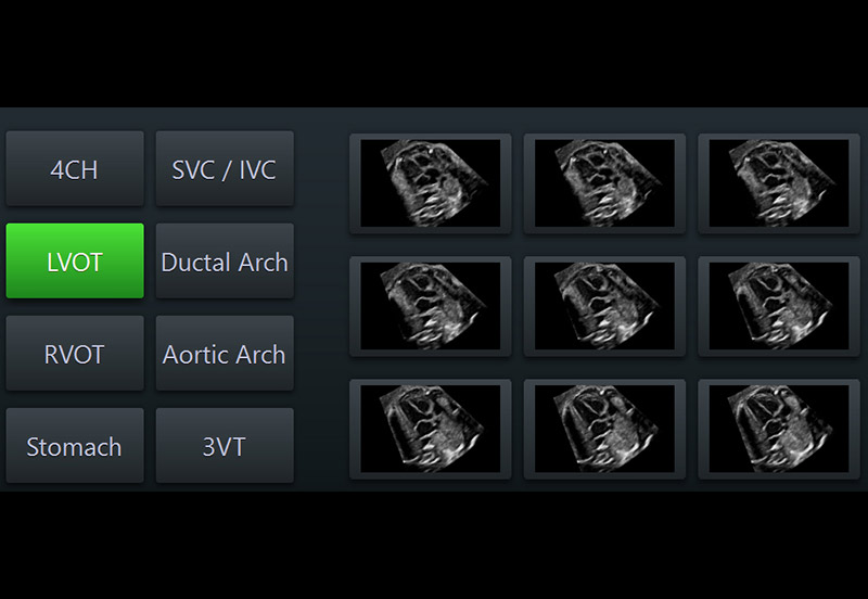
SonoVCAD™heart II - Display of LVOT view with a simple press of a touch screen button
With SonoVCAD™heart, OBGYNs and sonographers can easily visualize the anatomical structures in the International Society of Ultrasound in Obstetrics and Gynecology Practice Guidelines for the sonographic screening examination of the fetal heart.² The Voluson™ feature standardizes orientation of the heart and provides the recommended views obtained from a single volume or STIC* acquisition.
While SonoVCAD™heart is a huge step in making the exam easier and more accessible to physicians, Abuhamad is pushing for more innovation. He says GE is on the verge of creating revolutionary technology that would simplify the fetal heart exam even more. To accomplish Abuhamad’s vision, users would only need to make one decision—normal or abnormal.
“I’ve been in this business 25 years and I have been really amazed at our ability and the GE engineers’ ability to make ultrasound better and better, but this is only the tip of the iceberg,” Abuhamad said.
With each advancement, comes more opportunity to make an impact. And in the case of congenital heart disease, you can improve outcomes by identifying abnormalities sooner. Research shows that a prenatal diagnosis can significantly reduce preoperative and postoperative mortality rates for certain fetal heart abnormalities.³,⁴
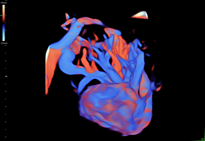
STIC acquisition highlighted with Voluson HDlive™ Flow for vessel detail
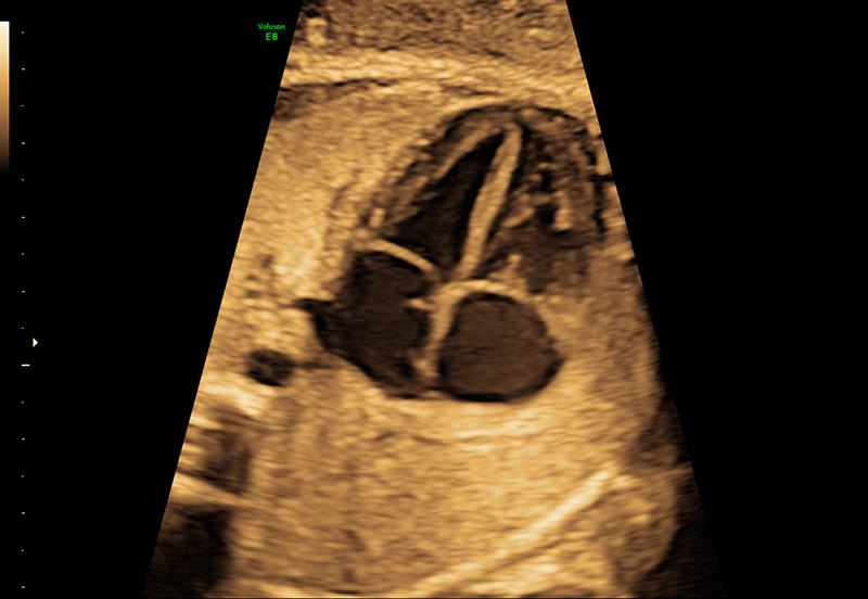
The 4-chamber view
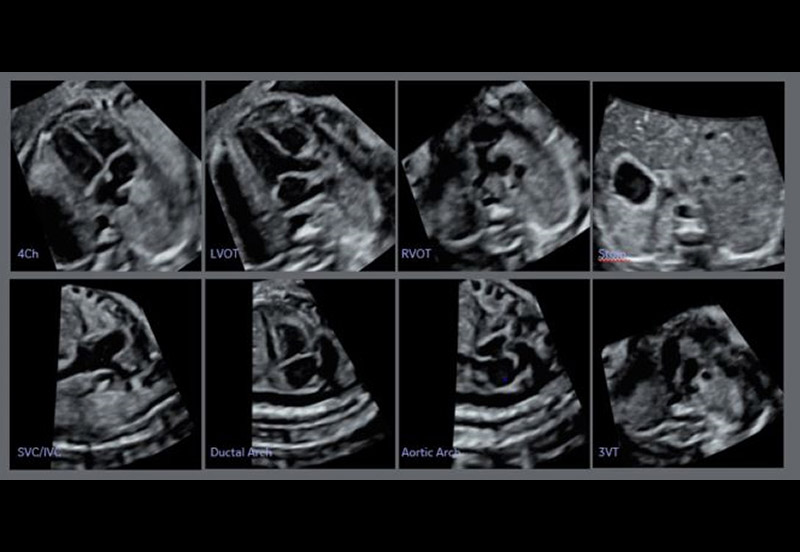
Correctly aligned fetal heart in multiplanar view
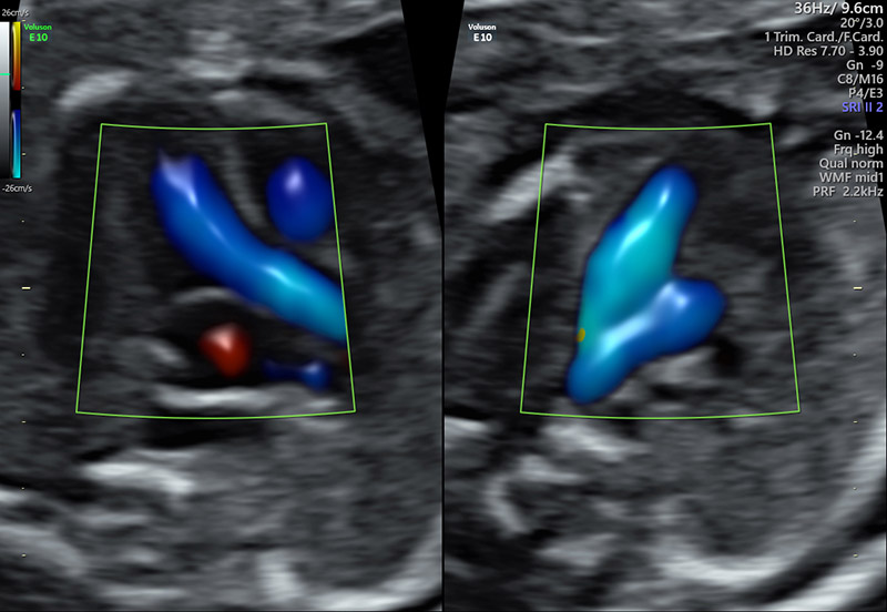
Dual display of the 3 Vessel view and LVOT with Voluson™ Radiantflow
“It’s a very stressful time for patients. Our ability to make the diagnosis early so we can privately counsel, support and plan the management of the baby after birth to improve the outcome—is really important,” Abuhamad stressed. “Creating tools that can get to that level is incredibly powerful.”
*Sonography-based Volume Computer Aided Display heart
*Spatio-Temporal Image Correlation
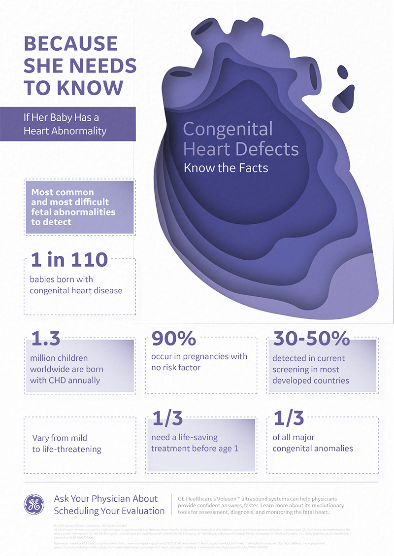
Download a free poster detailing some of the most important facts about fetal heart abnormalities for your examination room or office. Can be sized to 33.1h x 23.4w. (For best results, send to a professional printer.)
-
Learn more about how Voluson Sono-Automation technology can help simplify the fetal heart assessment and help provide answers, faster..
-
View Dr. Abuhamad’s ISUOG lecture ‘Towards full automation of the fetal heart examination’.
-
Explore here how the Voluson E10’s innovative technologies can help you focus on early prevention.
-
Explore here how Voluson 3D Printing for clinical prototyping can support your research efforts and enhance communication with your patient around fetal heart abnormalities.

