

It’s considered one of the most common and complex problems in modern obstetrics, with approximately 30 million newborns diagnosed with intrauterine growth restriction every year.¹ But a major advancement in ultrasound could help you get ahead of it.
Diagnosing Intrauterine Growth Restriction with Ultrasound
Signaling IUGR Sooner
Read how ultrasound technology is making it easier to diagnose intrauterine growth restrictions.

It’s considered one of the most common and complex problems in modern obstetrics, with approximately 30 million newborns diagnosed with intrauterine growth restriction every year.¹ But a major advancement in ultrasound could help you get ahead of it.
Some babies are just small. But others have problems developing because of intrauterine growth restrictions. It’s a serious condition that can be difficult to diagnose, but revolutionary new fetal heart technology from GE Healthcare is making it easier to determine which fetuses could be in danger.
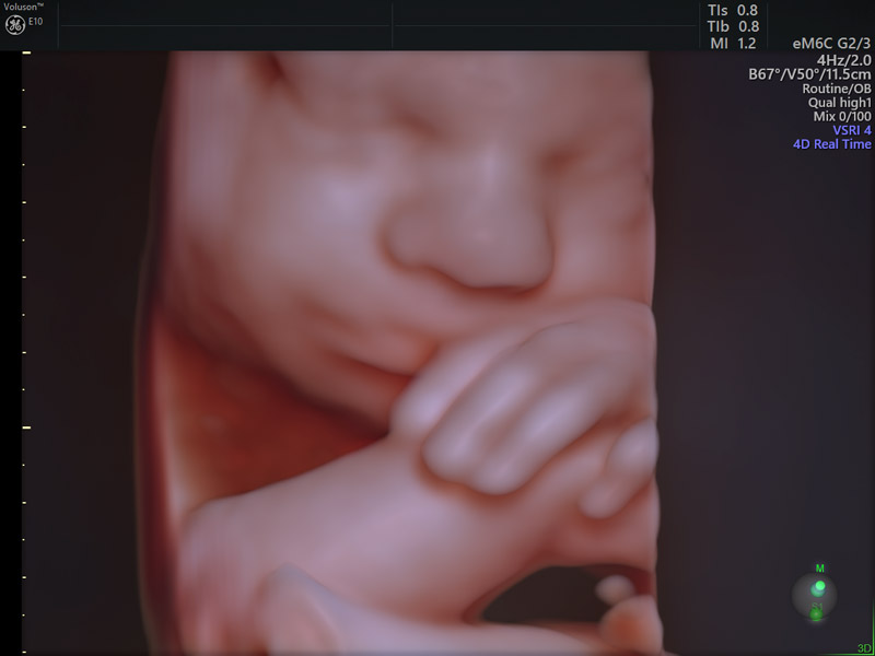
3D ultrasound rendering
The innovative tool is called fetalHQ and is featured on Voluson™ E10 ultrasound systems. For the first time, it’s possible to simultaneously examine the size, shape and contractility of the fetal heart from the 4-chamber view, based on 2D imaging and speckle tracking.
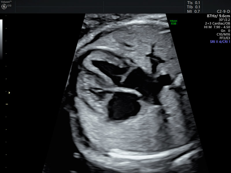
4-chamber view of fetal heart
The man behind the breakthrough is Greggory DeVore, M.D., a specialist in maternal fetal medicine and clinical Professor of Obstetrics and Gynecology at the David Geffen School of Medicine at UCLA in southern California. He came up with the idea and modified existing software designed to measure pediatric and adult hearts. DeVore then published 13 peer-reviewed articles that describe the clinical value of the new assessment and make up the backbone of fetalHQ.²⁻¹⁵
“For 50 years, we’ve only used heart rate patterns to look at cardiac function, but we now know what’s happening when the heart contracts with each beat. With fetalHQ, we can better visualize the changes in function, shape and size of the heart. That information gives us clues about the fetal heart before the fetus is in trouble,” DeVore explained.
The most common definition for IUGR is a fetal weight that is below the 10th percentile for gestational age. But making the clinical diagnosis can be a challenge when you are trying to scan a fetus that is in constant motion. It takes skill to measure blood flow in the umbilical cord and vessels in the baby’s brain, as well as heart function using Doppler tests.
“It’s very time consuming—and you’re not getting all the information you need,” DeVore said. “fetalHQ is easier to use, and you can collect all of the information in less than three minutes. We can analyze more parameters, so we can pick up more things that are wrong.”
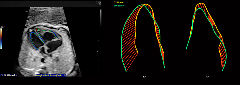
fetalHQ
Dr. DeVore points to his research on fetuses with different degrees of growth restriction. He found cases where fetuses had normal Dopplers, but their hearts were abnormal in size, shape, and function.
“The Doppler was okay, but their hearts were bad,” DeVore said. “If something is going wrong, the heart is the first thing that changes because it tries to adapt. The fetalHQ tool can help you see changes in heart function before the fetus develops IUGR.”
The ability to assess risk and identify problems earlier could ultimately lead to better outcomes. In fact, research shows timely diagnosis and management of IUGR can reduce perinatal mortality.¹⁶,¹⁷ DeVore believes Voluson’s fetalHQ will make a huge impact—not just in signaling IUGR, but anticipating other complications too.
“I believe that where we’re going with this, we’ll be able to predict which fetus gets into trouble in labor before it happens. I’ll be able to say, ‘the fetal heart is not working properly’ before the baby is in distress. That’s a very big deal.”
-
Learn more about Voluson fetalHQ – download abstracts.
-
Explore here how the Voluson E10’s innovative technologies can help you focus on early prevention.
-
Download a free poster detailing some of the most important facts about fetal heart abnormalities for your examination room or office. Can be sized to 33.1h x 23.4w.
-
Learn more about how Voluson Sono-Automation technology can help simplify the fetal heart assessment and help provide answers, faster.
-
Explore here how Voluson 3D Printing for clinical prototyping can support your research efforts and enhance communication with your patient around fetal heart abnormalities.
https://findinganswers.volusonclub.net/downloads/2018_Voluson%20Expert_BT19_Fetal_Heart_dedicated_sales_piece.pdf
DeVore GR, Tabsh K, Polanco B, Satou G, Sklansky M. Fetal Heart Size: A Comparison Between the Point-to-Point Trace and Automated Ellipse Methods Between 20 and 40 Weeks' Gestation. J Ultrasound Med. 2016 Dec;35(12):2543-2562.
DeVore GR, Polanco B, Satou G, Sklansky M. Two-Dimensional Speckle Tracking of the Fetal Heart: A Practical Step-by-Step Approach for the Fetal Sonologist. J Ultrasound Med. 2016 Aug;35(8):1765-81.
DeVore GR, Satou G, Sklansky M. Area of the fetal heart's four-chamber view: A practical screening tool to improve detection of cardiac abnormalities in a low-risk population. Prenat Diagn. 2017 Feb;37(2):151-155.
DeVore GR. Computing the Z Score and Centiles for Cross-sectional Analysis: A Practical Approach. J Ultrasound Med. 2017 Mar;36(3):459-473.
DeVore GR, Satou G, Sklansky M. Abnormal Fetal Findings Associated With a Global Sphericity Index of the 4-Chamber View Below the 5th Centile. J Ultrasound Med. 2017 Nov; 36(11): 2309-2318
DeVore GR, Klas B, Satou G, Sklansky M. Evaluation of the right and left ventricles: An integrated approach measuring the area, length, and width of the chambers in normal fetuses. Prenat Diagn. 2017 Dec;37(12):1203-1212.
DeVore GR, Klas B, Satou G, Sklansky M. 24-segment sphericity index: a new technique to evaluate fetal cardiac diastolic shape. Ultrasound Obstet Gynecol. 2018 May;51(5):650-658
DeVore GR, Klas B, Satou G, Sklansky M. Longitudinal Annular Systolic Displacement Compared to Global Strain in Normal Fetal Hearts and Those With Cardiac Abnormalities. J Ultrasound Med. 2018 May;37(5):1159-1171.
DeVore GR, Klas B, Satou G, Sklansky M. Twenty-four Segment Transverse Ventricular Fractional Shortening: A New Technique to Evaluate Fetal Cardiac Function. J Ultrasound Med. 2018 May;37(5):1129-1141.
DeVore GR, Zaretsky M, Gumina DL, Hobbins JC. Right and left ventricular 24-segment sphericity index is abnormal in small-for-gestational-age fetuses. Ultrasound Obstet Gynecol. 2018 Aug;52(2):243-249.
DeVore GR, Klas B, Satou G, Sklansky M. Quantitative Evaluation of the Fetal Right and Left Ventricular Fractional Area Change Using Speckle Tracking Technology. Ultrasound Obstet Gynecol. 2018 Mar 14. doi: 10.1002/uog.19048. [Epub ahead of print] PubMed PMID: 29536575.
DeVore GR, Klas B, Satou G, Sklansky M. Speckle Tracking of the Basal Lateral and Septal Wall Annular Plane Systolic Excursion of the Right and Left Ventricles of the Fetal Heart. J Ultrasound Med. 2018 Sep 12. doi: 10.1002/jum.14811. [Epub ahead of print] PubMed PMID: 30208238.
DeVore GR, Klas B, Satou G, Sklansky M. Evaluation of Fetal Left Ventricular Size and Function Using Speckle-Tracking and the Simpson Rule. J Ultrasound Med. 2018 Sep 23. doi: 10.1002/jum.14799. [Epub ahead of print] PubMed PMID: 30208238.
Manning FA, Hohler C. Intrauterine growth retardation: diagnosis, prognostication, and management based on ultrasound methods. In: Fleischer AC, et al., eds. The principles and practice of ultrasonography in obstetrics and gynecology. 4th ed. Norwalk, Conn.: Appleton & Lange, 1991:331–48.
Craigo SD. The role of ultrasound in the diagnosis and management of intrauterine growth retardation. Semin Perinatol. 1994;18:292–304.

