

Between 15 and 20% of identical twins that share the same placenta are affected by twin to twin transfusion syndrome.¹ It can lead to life-threatening complications and developmental defects—including intrauterine growth restriction, cardiomyopathy and neurologic impairment.
How Ultrasound Helps Make Critical Decisions in the Treatment of TTTS
A New Take on a Troubling Condition That Affects Identical Twins
Learn how fetalHQ, the latest ultrasound technology from GE Healthcare, could impact critical decisions in treating your patients with TTTS.

Between 15 and 20% of identical twins that share the same placenta are affected by twin to twin transfusion syndrome.¹ It can lead to life-threatening complications and developmental defects—including intrauterine growth restriction, cardiomyopathy and neurologic impairment.
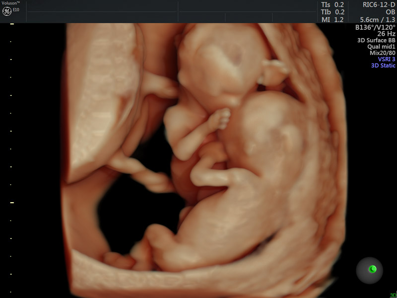
Twin pregnancy in single amniotic sac
Three minutes is all you need. A revolutionary new tool quickly collects vital information about the fetal heart—starting as early as 20 weeks. And what you discover could impact critical decisions in treating your patients with twin to twin transfusion syndrome.
While its speed is impressive, that’s not the most remarkable thing about fetalHQ—the latest ultrasound technology from GE Healthcare. The feature on the Voluson™ E10 systems measures the heart in ways that have never been done before. Clinicians can now determine the size, shape, and contractility of the heart from the 4-chamber view, based on 2D imaging and speckle tracking.
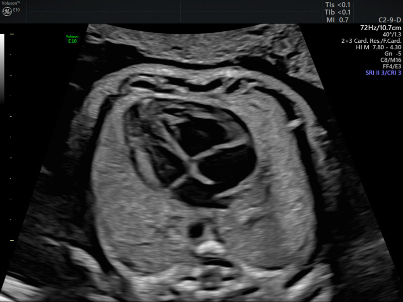
4-chamber view of fetal heart
“This tool is simple and sophisticated. It’s going to provide us with a new understanding of the fetal heart,” said Greggory DeVore, M.D., a specialist in maternal fetal medicine and clinical Professor at the David Geffen School of Medicine at UCLA in southern California.
Dr. DeVore came up with the idea and adapted existing software for pediatric and adult hearts to create the advanced fetal heart technology. He published 13 peer-reviewed articles with clinical data that serve as the foundation for fetalHQ.²-¹⁵ DeVore knew that with more information about fetal heart function, physicians could identify abnormalities, and possibly get ahead of life-threatening prenatal conditions like intrauterine growth restriction. He also sees great potential to better understand and treat twin to twin transfusion syndrome.
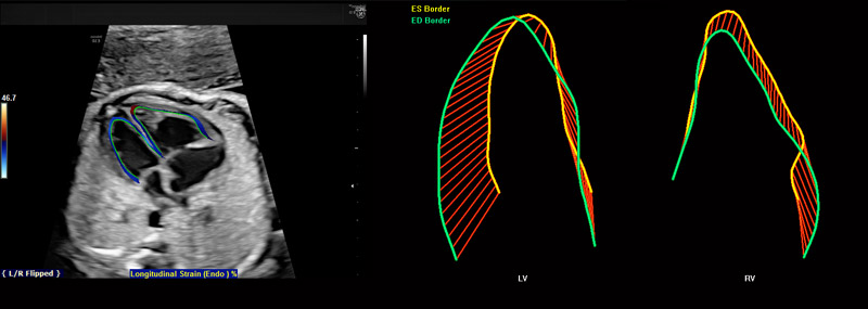
fetalHQ
“We want to know how the hearts are functioning, and think fetalHQ could be a great tool to really dissect cardiac function in both fetuses,” DeVore attested. “With more assessment, we’ll ask the question—should we intervene earlier or later? Maybe it will help give us an answer.”
If left untreated, TTTS has a 90% mortality rate—and the babies that survive can face significant challenges. There is a 30 to 50% risk of neurological problems including cerebral palsy, vision loss, and hearing loss.¹⁶ ¹⁷
Ultrasound already plays an important role in making critical decisions in the treatment of TTTS. Evaluations are often based on the weight of the fetuses, amount of amniotic fluid, and how well blood is flowing between babies using umbilical artery Doppler studies. The most common treatment is fetoscopic laser photocoagulation—which is reserved for advanced cases of TTTS, when the situation is dire.
“What we’re trying to do is fine-tune the analysis,” DeVore said. “We’re just getting into this, but I think at some point we’ll be able to ask, ‘Are the changes in the heart such that we should do laser therapy earlier?’ With time and expertise, maybe we’d have a much better outcome.”
DeVore believes the Voluson tool will lead to more questions, and ultimately more insight. “With fetalHQ, we can ask all kinds of questions—and once we know how the puzzle fits together, we can understand the big picture. We think it’s going to revolutionize fetal care.”
-
Learn more about Voluson fetalHQ – download abstracts.
-
Explore here how the Voluson E10’s innovative technologies can help you focus on early prevention.
-
Download a free poster detailing some of the most important facts about fetal heart abnormalities for your examination room or office. Can be sized to 33.1h x 23.4w.
-
Learn more about how Voluson Sono-Automation technology can help simplify the fetal heart assessment and help provide answers, faster.
-
Explore here how Voluson 3D Printing for clinical prototyping can support your research efforts and enhance communication with your patient around fetal heart abnormalities.
Sebire NJ, Snijders RJM, Hughes K, Sepulveda W, Nicolaides KH. The hidden mortality of monochorionic twin pregnancies. Br J Obstet Gynaecol 1997;104:1203-7.
DeVore GR, Tabsh K, Polanco B, Satou G, Sklansky M. Fetal Heart Size: A Comparison Between the Point-to-Point Trace and Automated Ellipse Methods Between 20 and 40 Weeks' Gestation. J Ultrasound Med. 2016 Dec;35(12):2543-2562.
DeVore GR, Polanco B, Satou G, Sklansky M. Two-Dimensional Speckle Tracking of the Fetal Heart: A Practical Step-by-Step Approach for the Fetal Sonologist. J Ultrasound Med. 2016 Aug;35(8):1765-81.
DeVore GR, Satou G, Sklansky M. Area of the fetal heart's four-chamber view: A practical screening tool to improve detection of cardiac abnormalities in a low-risk population. Prenat Diagn. 2017 Feb;37(2):151-155.
DeVore GR. Computing the Z Score and Centiles for Cross-sectional Analysis: A Practical Approach. J Ultrasound Med. 2017 Mar;36(3):459-473.
DeVore GR, Satou G, Sklansky M. Abnormal Fetal Findings Associated With a Global Sphericity Index of the 4-Chamber View Below the 5th Centile. J Ultrasound Med. 2017 Nov; 36(11): 2309-2318
DeVore GR, Klas B, Satou G, Sklansky M. Evaluation of the right and left ventricles: An integrated approach measuring the area, length, and width of the chambers in normal fetuses. Prenat Diagn. 2017 Dec;37(12):1203-1212.
DeVore GR, Klas B, Satou G, Sklansky M. 24-segment sphericity index: a new technique to evaluate fetal cardiac diastolic shape. Ultrasound Obstet Gynecol. 2018 May;51(5):650-658
DeVore GR, Klas B, Satou G, Sklansky M. Longitudinal Annular Systolic Displacement Compared to Global Strain in Normal Fetal Hearts and Those With Cardiac Abnormalities. J Ultrasound Med. 2018 May;37(5):1159-1171.
DeVore GR, Klas B, Satou G, Sklansky M. Twenty-four Segment Transverse Ventricular Fractional Shortening: A New Technique to Evaluate Fetal Cardiac Function. J Ultrasound Med. 2018 May;37(5):1129-1141.
DeVore GR, Zaretsky M, Gumina DL, Hobbins JC. Right and left ventricular 24-segment sphericity index is abnormal in small-for-gestational-age fetuses. Ultrasound Obstet Gynecol. 2018 Aug;52(2):243-249.
DeVore GR, Klas B, Satou G, Sklansky M. Quantitative Evaluation of the Fetal Right and Left Ventricular Fractional Area Change Using Speckle Tracking Technology. Ultrasound Obstet Gynecol. 2018 Mar 14. doi: 10.1002/uog.19048. [Epub ahead of print] PubMed PMID: 29536575.
DeVore GR, Klas B, Satou G, Sklansky M. Speckle Tracking of the Basal Lateral and Septal Wall Annular Plane Systolic Excursion of the Right and Left Ventricles of the Fetal Heart. J Ultrasound Med. 2018 Sep 12. doi: 10.1002/jum.14811. [Epub ahead of print] PubMed PMID: 30208238.
DeVore GR, Klas B, Satou G, Sklansky M. Evaluation of Fetal Left Ventricular Size and Function Using Speckle-Tracking and the Simpson Rule. J Ultrasound Med. 2018 Sep 23. doi: 10.1002/jum.14799. [Epub ahead of print] PubMed PMID: 30208238.
Saunders NJ, Snijders RJM, Nicolaides KH. Therapeutic amniocentesis in twin-twin transfusion syndrome appearing in the second trimester of pregnancy. Am J OBstet Gynecol 1992;166:820-4.
Haverkamp F, Lex C, Hanisch C, Fahnenstich H, Zerres K. Neurodevelopmental risks in twin-to-twin transfusion syndrome: preliminary findings. Eur J Pediatric Neurology 2001:5:21-7.

