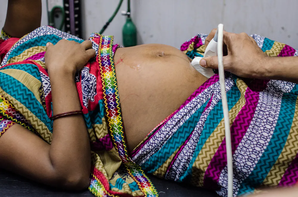

Prenatal ultrasound is used throughout pregnancy to evaluate fetal health. Ultrasound of the fetal heart is only one component of standard prenatal screening, but it has the potential to greatly improve outcomes for babies born with congenital heart defects (CHDs). Dr. Balu Vaidyanathan, a pediatric cardiologist in Kerala, India, is focused on this problem and seeing real results.
The Scope of Congenital Heart Disease
The Centers for Disease Control and Prevention names CHDs as the most common of all birth defects. CHDs caused more than 9 million deaths in 2016 in children under five years old, according to the Annals of Public Health Reports. In India, where Dr. Balu practices, approximately 240,000 babies per year are born with a heart defect.
eSTIC (electronic Spatio-Temporal Image Correlation) – Enhances fetal cardiac exams with up to 75% reduction in acquisition time over traditional STIC and delivers improved resolution in the B and C planes**
Although CHDs are a major cause of morbidity and mortality around the world, diagnosing them during pregnancy can have a significant positive impact. Dr. Balu credits the use of high-definition ultrasound to diagnose CHDs prenatally as one factor that enabled the Indian state of Kerala to reduce its infant mortality rate by three per 1,000 births. That change represents thousands of lives saved.
Dr. Balu points out that the best way to care for babies with CHDs is to establish a prenatal diagnosis so the baby can receive appropriate care immediately after birth. Because CHD treatment can range from a "wait and see" approach to surgical intervention, a prenatal ultrasound offers critical foresight into an infant's future needs.
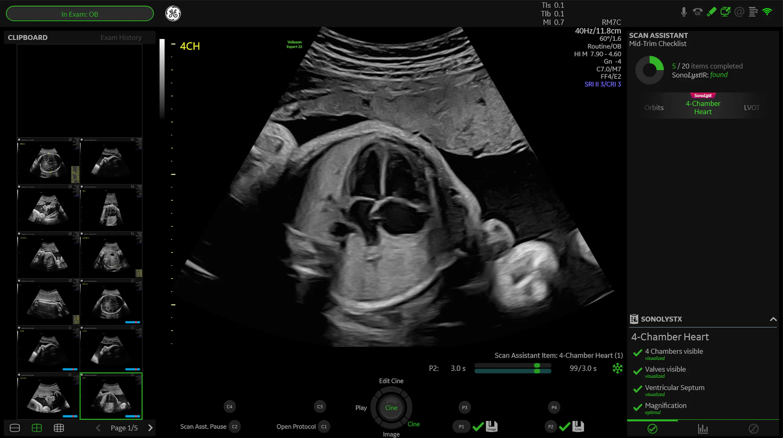
AI technology: SonoLyst* – Automatically identifies and annotates standard second-trimester fetal heart views to enhance efficiency – compare acquired image/view to standard criteria for quality assurance, ensuring image quality and consistency.
Dr. Balu notes the impact of prenatal diagnosis particularly when caring for patients who live in an area without a pediatric cardiac facility. Heart defects in newborns may not be detected during routine screening at birth, so they may go home from the hospital undiagnosed — only to become severely ill later. Other children will be too sick to survive after delivery without immediate treatment. After receiving a prenatal diagnosis, expectant parents are better equipped to make arrangements to deliver in a facility that can provide the necessary treatment for their child.
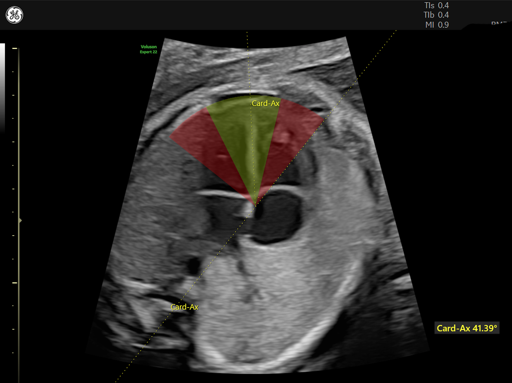
Cardiac Axis is one of the automated measurements in the fetalHS tool, to help quickly identify the potential for a congenital heart defect.
Children who receive surgical intervention earlier are not only surviving at very high rates (98% in Dr. Balu's facility), but they are also spending fewer days in the hospital, as Dr. Balu has witnessed firsthand.
With the increase in tertiary care centers in India and growing availability of ultrasound services, Dr. Balu sees the life-saving potential that comes with being able to diagnose more children with CHDs prenatally. Although the International Society Ultrasound in Obstetrics and Gynecology calls for second-trimester screening of the heart's anatomy, Dr. Balu and other providers are successfully diagnosing many CHDs in the first trimester.
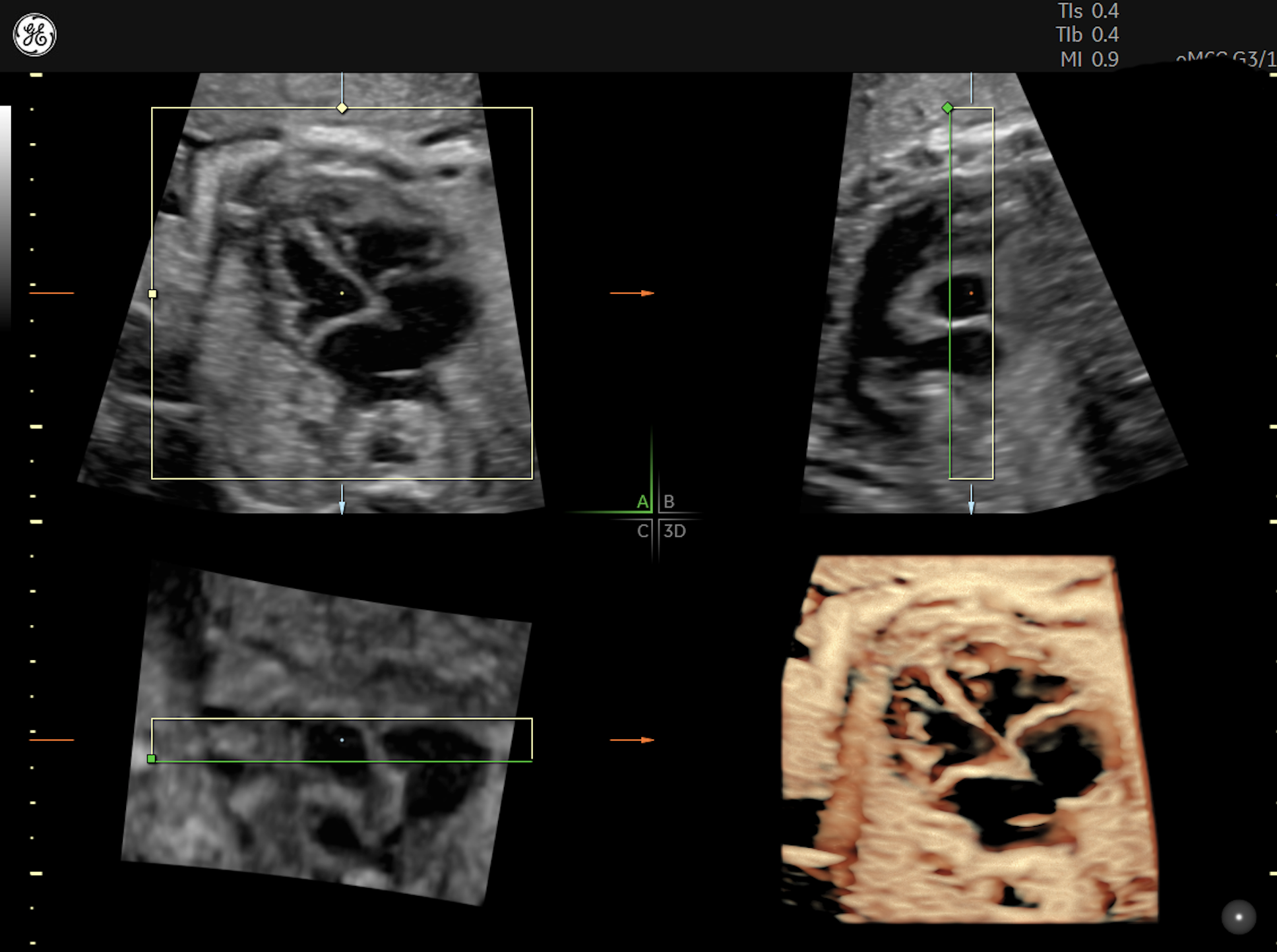
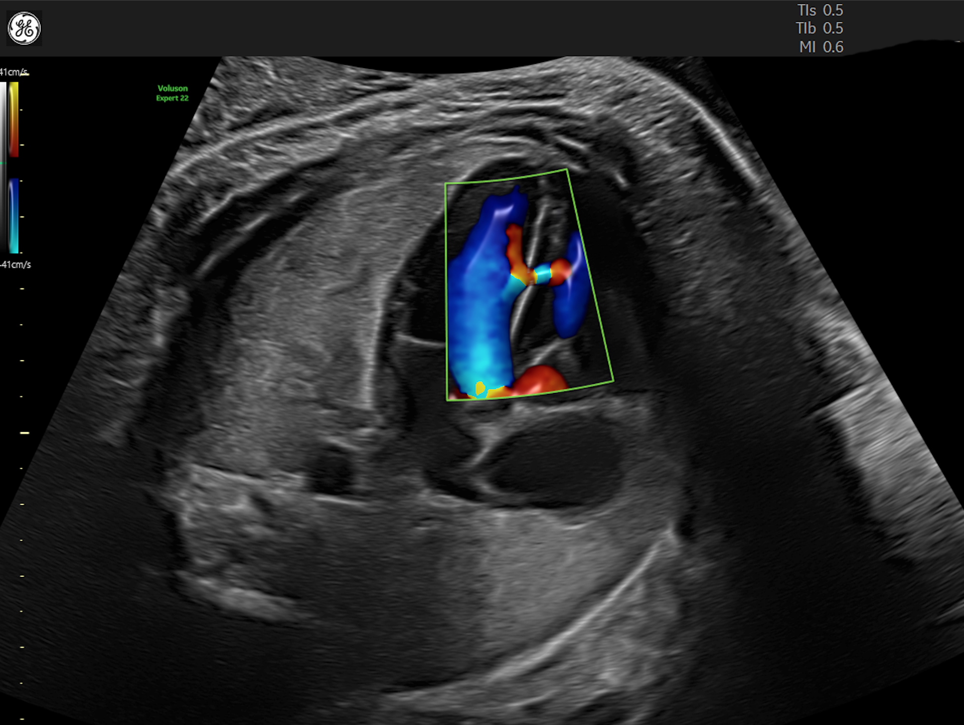
Early detection of CHDs on ultrasound relies heavily on two things — operator skill and the right equipment. Dr. Balu uses the Voluson E10, which uses artificial intelligence to quickly produce high-quality images from 2D and 3D while reducing the number of steps required by the technician. Using color Doppler during these exams gives extra insight into blood flow through the cardiac anatomy.

e4D SnapShot – Optimizes exam time with one-button access from Real-Time 4D to acquire an eSTIC data set.
As Translational Pediatrics explains, screening for CHDs no longer relies solely on the standard four-chamber view, because it can miss a number of cardiac anomalies. Also, imaging the ventricular outflow tracts has been recommended since approximately 2013.
Translational Pediatrics recommends that suspected CHDs be further evaluated by fetal echocardiography using 2D imaging, Doppler (both color and pulse-wave), and M-mode. The transducer frequency should be between 4 and 12 MHz. The complete exam should include still and moving images of the heart itself, flow through the valves and flow in and out of the heart, paying special attention to any abnormal flows. Additionally, the cardiac chambers, valve annuli and vessels should be measured.
Dr. Balu further enhances his prenatal cardiac studies by employing spatio-temporal image correlation (STIC) using 3D/4D imaging to provide detailed metrics of the cardiac anatomy, which aids in more precise surgical planning. The Journal of Cardiovascular Imaging reinforces the value of STIC imaging, noting its capability to assess valve function and cardiac volume through a complete cardiac cycle.
Physicians like Dr. Balu, who use prenatal ultrasound to diagnose CHDs, witness the heartbreak and worry that come with this diagnosis. Yet, he is also able to help parents see the success of his efforts, as more and more babies thrive after receiving appropriate care.
Radiantflow™ delivers easy, fast visualization of blood flow using the amplitude of color Doppler signal to enhance the robustness and create a 3D-like appearance.
*SonoLyst incorporates the technology of Intelligent Ultrasound