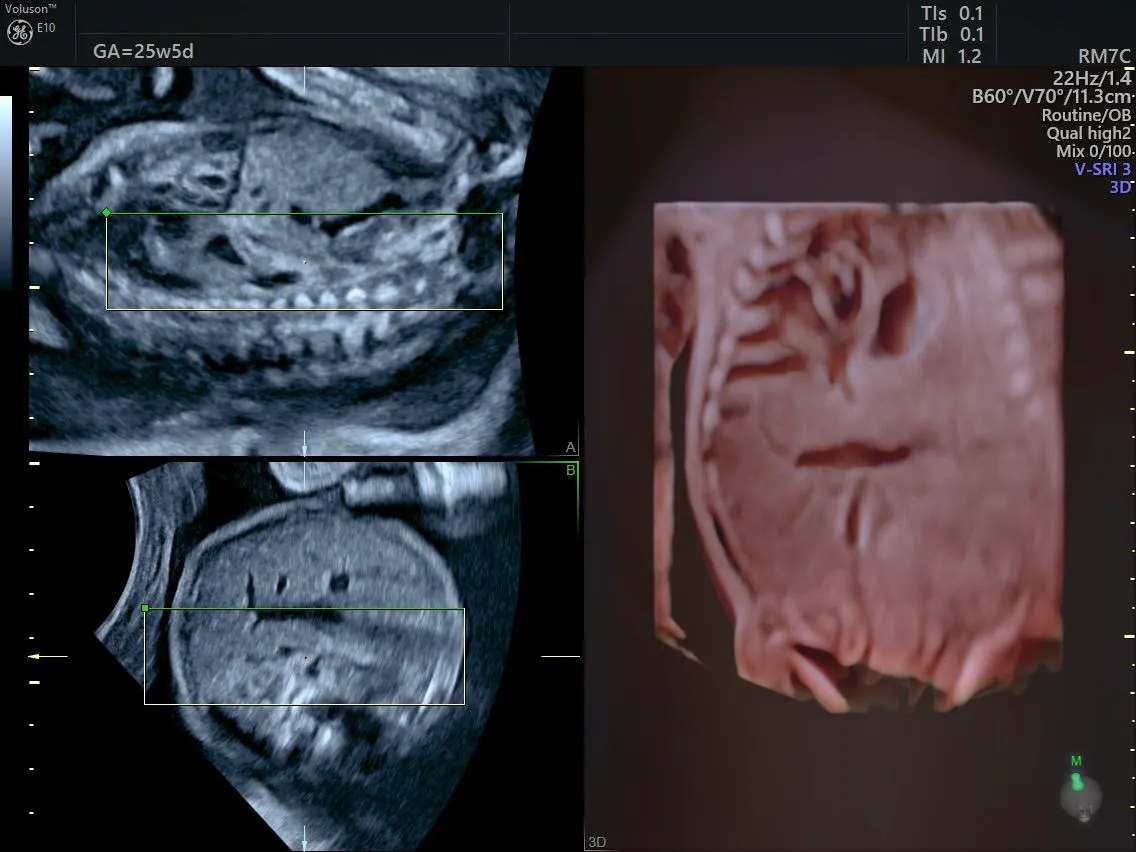
Diagnosing Fetal Diaphragmatic Hernia With Ultrasound
A fetal diaphragmatic hernia occurs when there is a defect or a weakness in the muscle of the fetal diaphragm, which divides the abdominal and thoracic cavities. This defect can occur on just one side or bilaterally, on both the left and right sides. However, it most commonly affects the left side of the diaphragm.
When this defect occurs, the organs of the abdomen can herniate through into the thorax, impacting the fetus's lung development. This can potentially lead to pulmonary hypoplasia, a condition that affects not only breathing but also heart function and overall development.
Diaphragmatic hernias are rare conditions that affect about 1 in every 3,600 babies born in the US, reports the Centers for Disease Control and Prevention. Further, approximately 50 percent of babies born with a diaphragmatic hernia can also have other significant congenital defects, says a study published by the National Library of Medicine. The exact cause of this condition is not yet fully understood; however, current theories point to abnormalities in the genes.
Diagnosing Diaphragmatic Hernia Fetal Ultrasound
Most cases of diaphragmatic hernia can be identified on obstetric ultrasound in the second or third trimester of pregnancy. This is done by assessing the abdominal and thoracic organs to determine if they are in normal positions relative to each other and the fetal diaphragm.
With a left-sided herniation of the stomach through the diaphragm, ultrasound can show whether the heart has shifted to the right and the stomach bubble is at the same level as the heart. This situation is the easiest fetal diaphragmatic hernia to identify. Other forms of hernia on the right side, or those that do not involve the stomach, can be more challenging to identify, as these organs appear similar in echotexture and echogenicity to lung tissue. However, direct visualization of the diaphragm in the sagittal plane can help identify these conditions. Fetal diaphragmatic hernia can also be identified through the use of first-trimester anomaly screening.
An article published in Prenatal Diagnosis reports that 25 percent of cases with this condition are associated with an increased nuchal translucency — a diagnosis that should lead to a detailed second-trimester obstetric ultrasound for confirmation of abnormalities. Prenatal Diagnosis also says that in nearly 50 percent of cases, a fetal diaphragmatic hernia is found alongside other structural anomalies and that these cases can be indicative of syndromes or chromosomal disorders.
There are more than 100 genetic syndromes that may include a fetal diaphragmatic hernia. The most common and concerning structural anomaly is abnormalities of the fetal heart, which occur in 50 percent of cases. There can also be anomalies in the brain, kidneys, bones and spine in 10 percent of these cases. Advanced ultrasound imaging should be relied on to determine if any other structural anomalies are present in a fetus with a known diaphragmatic hernia.
Managing and Treating Fetal Diaphragmatic Hernia
Depending on the total number of anomalies affecting the fetus, the parents are given the option of either expectant management, termination of pregnancy or fetal intervention.
- Expectant management is a follow-up similar to that of a normal uncomplicated pregnancy.
- Termination may be considered in severe cases with poor expected outcomes.
- Fetal intervention can be done in certain situations to improve the chances for the baby after delivery. For example, surgical intervention can be done for fetuses with moderate to severe pulmonary hypoplasia, where a balloon is inserted into the fetal trachea to trap secretions from the lungs, promoting stretch-induced lung growth. This is done through a fetoscopic endoluminal trachea occlusion procedure, which is minimally invasive.
Delivery of a fetus affected by a diaphragmatic hernia is planned at a surgical center to allow for the best surgical options and care after birth. Intensive management of the baby at birth is required, which can include: intubation and ventilation, sedation, and drugs that improve circulation and treat pulmonary hypertension.
After the baby is stabilized after birth, they are scheduled for a surgical repair of the diaphragmatic hernia and repositioning of the herniated organs back into the abdominal space. This can be done via laparotomy or laparoscopy through the abdomen or via thoracotomy or thoracoscopy through the thorax.
Providing Effective Care and Communication
To best counsel and provide care to obstetric patients learning about a diaphragmatic hernia affecting their neonate, clinicians need to provide clear communication and information about the condition, as well as thorough explanations of the treatment options available.
A detailed and targeted diaphragmatic hernia fetal ultrasound exam should also be completed and explained to the parents, giving them time to digest the information and ask questions about next steps. In this way, clinicians can provide the most effective care, support and guidance.

