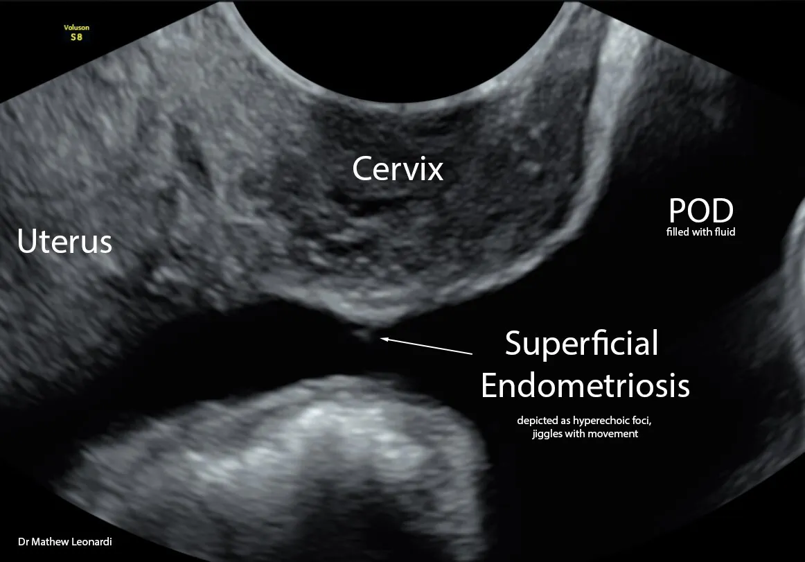
Diagnosis of Endometriosis, Including Superficial Endometriosis, Using Ultrasound
Endometriosis is a puzzling and long-lasting inflammatory condition in which tissue similar to the lining of the uterus grows outside of it. The diagnosis of endometriosis is of the utmost importance: those affected experience debilitating pain during periods, pain during urination, discomfort during sex, and difficulties getting pregnant. In some cases, endometriosis can show up in unexpected places, like the lung, liver, or spleen.
There are three main types of endometriosis, each with its own characteristics:
1.Ovarian endometriomas: dark fluid-filled cysts in the ovaries
2.Deep endometriosis: invasion into nearby organs, like the bowel and bladder
3.Superficial endometriosis: thin lesions on the wall of the pelvis or abdomen
More specifically, superficial endometriosis (SE) may be marked by different colors, like black, red, white, or clear, depending on factors like blood supply and tissue changes. There's an ongoing discussion about whether endometriosis worsens over time; the color of the lesion, changing from red to black and eventually white or clear, might indicate its age.
Endometriosis depends on hormonal activity, particularly estrogen. The tissues involved show much higher levels of estrogen receptors compared to normal tissue, and hormones like 17β-Estradiol encourage the growth, inflammation, and pain associated with endometriosis. Interestingly, superficial endometriosis lesions can produce their own estrogen, compounding the problem. While the role of estrogen is quite clear, the role of progesterone in endometriosis remains the subject of a debate. In endometriosis, there's a lack of progesterone receptors, leading to progesterone resistance. Together, too much estrogen and this limited response to progesterone drive the disease, and traditional treatments aim to reduce estrogen or increase progesterone.
One significant problem—besides pain—caused by endometriosis, affecting around 30-50% of those with the condition, is infertility. The reasons for infertility in endometriosis can be clear (e.g., the distortion of pelvic anatomy seen in certain types of the condition) or less obvious (as with superficial endometriosis), but likely involve a mix of hormonal imbalances, molecular changes, and immune dysfunction that leave the uterus less receptive to a pregnancy. Specifically, for example, patients with endometriosis tend to have more prostaglandin, leading to abnormal contractions of the uterus in response to irritation and inflammation.
Advantages of Ultrasound as a Diagnostic Tool
An ultrasound diagnosis of endometriosis is a relatively affordable, quick, and non-invasive solution. Advances in transvaginal ultrasound (TVUS) have improved accuracy, making it a primary tool for investigating pelvic pain and other gynecological issues. In 2016, the International Deep Endometriosis Analysis (IDEA) group presented a consensus opinion standardizing ultrasound mapping for ovarian and deep endometriosis, enhancing its diagnostic value.
Recent reviews suggest that ultrasound is comparable to, and in some cases better than, laparoscopy for diagnosing endometriosis. Ultrasound has a sensitivity of 93% and specificity of 96% for ovarian endometriomas. When evaluating deep endometriosis using the IDEA group approach, accuracy depends on the lesion's location, mirroring laparoscopy results. Ultrasound excels in some areas, like predicting ASRM staging and utilizing the #ENZIAN classification system for specific compartments.
Clinicians have long believed that no imaging test, including ultrasound, could reveal superficial endometriosis. However, researchers are working to improve the sonographic accuracy of superficial endometriosis diagnosis. Initially, some looked for soft markers of the disease on ultrasound—visual features such as thickened uterosacral ligaments or ovarian immobility—but findings have been inconclusive. Others, who considered whether tenderness experienced by the patient during a TVUS could signify superficial endometriosis, met with similarly limited results.
A 2019 pilot study introduced a novel technique called saline infusion sonoPODography (SPG), an adaptation of the well-known saline infusion sonography that aims to visualize cases of superficial endometriosis. The procedure involves infusing the fluid into the pouch of Douglas (POD) through the uterus and Fallopian tubes via an intrauterine catheter, similar in technique to traditional sonohysterography (also known as SIS). In the majority of patients, open Fallopian tubes allows the fluid to move through and fill the POD, yielding a "standoff" view of posterior compartment structures, including uterosacral ligaments, torus uterinus, and the POD peritoneum.
Whilst SPG is still in its infancy, the study used particular descriptors to summarize the appearance of SE, relative to peritoneal surfaces, on TVUS:
1.Hyperechoic projections
2.Hypoechoic areas
3.Cystic areas
4.Filmy adhesions
5.Peritoneal pockets
In 2020, a second study followed up on this original publication by testing the procedure's accuracy in visualizing superficial endometriosis. When checking for SE in a general group of people with relevant signs and symptoms, it found, SPG can correctly identify cases about 69.1% of the time. The tool's sensitivity, or ability to correctly identify those with SE, is 64.9%. Its specificity, or ability to correctly identify those without SE, is 100%: SPG did not mistakenly diagnose superficial endometriosis in those unaffected.
However, when the study excluded patients with more severe types of endometriosis (ovarian and deep endometriosis) and obliteration of the POD, the accuracy of SPG improved. This is important: the most essential current diagnostic priority is to distinguish between populations with isolated superficial endometriosis and those with no endometriosis. In these cases, where only SE was considered, SPG showed an accuracy of 80%, with no false positives. This means it performed well in correctly identifying cases of isolated superficial endometriosis.
Critically, these results must be confirmed in larger studies to ensure that the technique works consistently across different groups of people. Further investigation is also necessary to understand how well people can tolerate this technique, what its limitations might be, and any associated pain experiences.
Challenges and Limitations
Despite the advancements detailed above, there are limitations to using ultrasound. Achieving high specificity and sensitivity requires extensive training and experience. Indeed, we may never gain the ability to rule out superficial endometriosis using ultrasound, or any imaging test, due to the usual very small size of some deposits. Even so, there's a need for increased awareness among healthcare providers and patients about the utility of non-invasive tools for endometriosis diagnosis.
As emphasis on ultrasound as a diagnostic tool increases, scholars and clinicians should further standardize terminology and adopt a step-wise approach beyond that resulting from the 2016 IDEA consensus statement. Unification of sonographic and surgical descriptors, particularly in superficial endometriosis, is necessary.
The Power of Ultrasound for Endometriosis Diagnosis
Ultrasound has already come a long way: it successfully diagnoses deep endometriosis and ovarian endometriomas and increasingly serves as a pre-operative surgical planning tool. Despite these innovations, superficial endometriosis, the most common subtype—and likely the earliest form of the disease—still evades ultrasound diagnosis. With advanced techniques, such as implementation of SPG or its lessons in the presence of natural peritoneal fluid, we can non-invasively detect this most subtle form of endometriosis for the first time in history. While there's more work to do, it is clear that the power of ultrasound continues to grow.
Interested in viewing Dr. Leonardi’s lecture, please click here: Ultrasound for superficial endometriosis.

