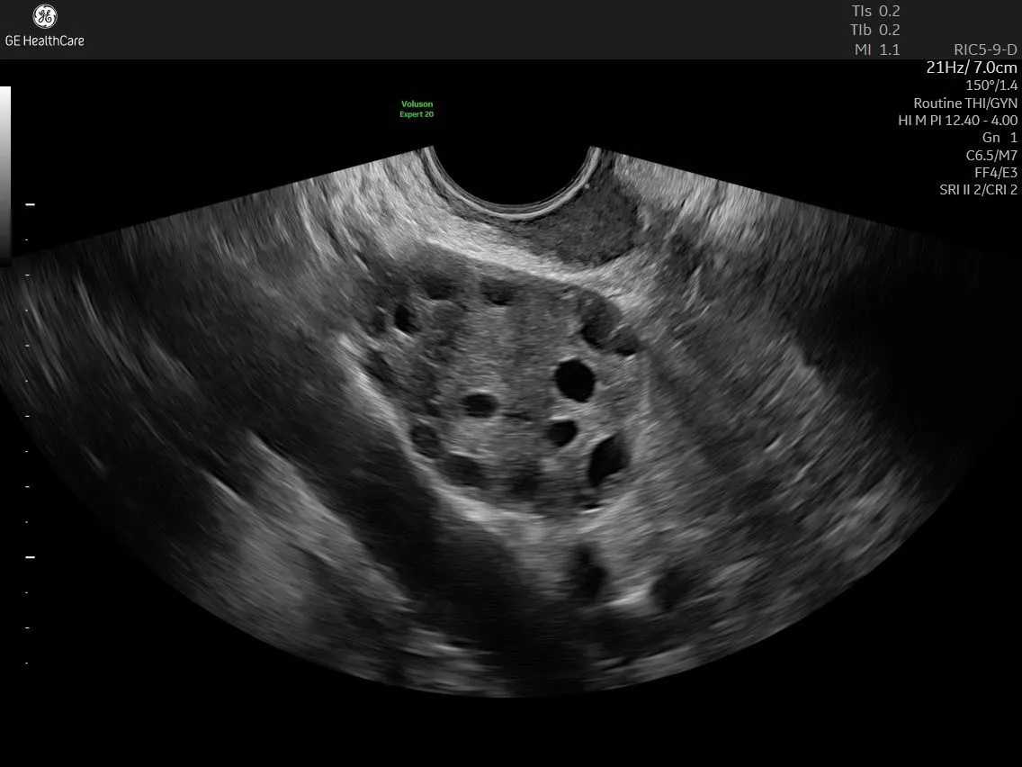
Earlier Recognition of PCOS and Endometriosis in Adolescents
Polycystic ovary syndrome (PCOS), also referred to as polyendocrine metabolic ovarian syndrome (PMOS), and endometriosis are often associated with adult women, yet both conditions can emerge during adolescence. Recognising the signs early can help clinicians support timely evaluation, facilitate appropriate management, and empower young patients to better understand their health.
As awareness of adolescent gynaecological health continues to grow, clinicians have an important opportunity to identify symptoms earlier, guide patients through the diagnostic journey, and help reduce the long-term impact these conditions can have on quality of life.
The Importance of Earlier Recognition
Despite increased awareness, diagnostic delays remain common. Studies suggest that women often experience delays of up to two years for PCOS and up to 10 years for endometriosis before receiving a diagnosis.¹˒²
Early recognition can be particularly challenging during adolescence. Irregular menstrual cycles are common in the years following menarche, painful periods may be perceived as normal, and gastrointestinal symptoms frequently overlap with those experienced throughout adolescence. In addition, many teenagers may feel uncomfortable discussing menstrual health, pelvic pain, constipation, or other sensitive symptoms.
PCOS and endometriosis can also present differently in adolescents than in adults. For this reason, clinicians benefit from understanding the nuances of adolescent presentations, recognising when further assessment may be appropriate, and using imaging and clinical evaluation to help build a complete picture of patient health.
Earlier evaluation and management can play an important role in supporting symptom control, reducing disease burden, and addressing emotional, social, and physical challenges that may accompany these conditions.³
Recognising Endometriosis in Adolescents
Pelvic pain remains the most common presenting symptom of endometriosis in adolescents. Dysmenorrhoea that limits daily activities, contributes to school absences, or fails to respond to first-line treatment should prompt further evaluation.⁴
Understanding the distinct characteristics of adolescent endometriosis can support earlier recognition. Compared with adults, adolescents are more likely to present with superficial peritoneal implants and are less likely to have large ovarian endometriomas.⁴ Deep infiltrating endometriosis (DIE) may still occur, particularly in patients with significant symptoms.
Laparoscopic findings in adolescents often differ from the classic appearance associated with adult disease. Lesions may appear clear, white, or as small haemorrhagic implants rather than the traditional dark "powder-burn" lesions. These early lesions are highly biologically active and may contribute significantly to pain symptoms.⁵
The Role of Ultrasound in Endometriosis Assessment
Ultrasound plays an important role in the assessment of adolescents with suspected endometriosis.
For adolescents who are not sexually active, transabdominal ultrasound serves as an appropriate first-line imaging approach. Ultrasound can help identify ovarian endometriomas, exclude alternative causes of pelvic pain, and document findings that may support referral for additional evaluation. However, it is important to recognise that a normal ultrasound examination does not exclude endometriosis.⁶
A systematic ultrasound assessment includes evaluation of the uterus, ovaries, bladder, vesicouterine space, uterosacral ligaments, rectovaginal septum, and rectosigmoid region. Clinicians should also assess for indirect signs of adhesive disease, including fixed ovaries, cul-de-sac obliteration, and reduced organ mobility during real-time imaging.⁶
Classic ovarian endometriomas typically appear as uni- or multilocular cysts with homogeneous low-level "ground-glass" echoes, smooth inner walls, and absent or minimal internal vascularity on Doppler imaging.⁵
Deep infiltrating endometriosis may present as hypoechoic nodules, thickening of the uterosacral ligaments, or involvement of the rectovaginal septum and rectosigmoid bowel wall.⁵ Abnormal ovarian positioning, fixed uterine retroversion, or tethering of bowel loops may provide additional clues, even when lesions are not directly visualised.⁷
A structured imaging protocol, including systematic documentation of both positive and negative findings, can strengthen diagnostic confidence and support comprehensive clinical decision-making.⁶
Evaluating PCOS in Adolescents
Assessment of PCOS during adolescence requires a different approach than in adults.
While the Rotterdam criteria are widely used in adult populations, their direct application during adolescence may increase the risk of overdiagnosis because multifollicular ovaries are commonly observed during normal pubertal development.
According to the 2023 International Evidence-Based Guideline for PCOS, adolescent diagnosis should include both hyperandrogenism and ovulatory dysfunction. Pelvic ultrasound and anti-Müllerian hormone (AMH) testing are not recommended as diagnostic criteria until eight years after menarche.¹
In adolescents, ultrasound primarily supports exclusion of alternative pathology rather than confirmation of PCOS. Imaging may be used to identify structural uterine or ovarian abnormalities, evaluate primary amenorrhoea, and rule out conditions such as ovarian masses, torsion, or congenital anatomical variants that require different management approaches.¹˒⁵
Current international guidance also recommends against using follicle number per ovary counts in patients younger than 18 years due to the high prevalence of multifollicular ovarian morphology during puberty.¹˒⁹
Clinical history, symptom assessment, physical examination, and laboratory findings remain essential components of adolescent PCOS evaluation. When appropriate, management may include lifestyle counselling, metabolic monitoring, and symptom-directed medical therapies. Ongoing screening for associated metabolic and psychological comorbidities is recommended throughout follow-up.¹
Supporting Adolescents Beyond Diagnosis
Successful care extends beyond the diagnostic process itself.
Adolescents benefit from being active participants in discussions regarding evaluation, treatment options, and long-term management alongside their parents or caregivers. Creating an environment that encourages open communication can help patients better understand their symptoms and become more engaged in their own healthcare journey.
With appropriate evaluation and symptom management, early recognition of PCOS and endometriosis can help improve quality of life, support mental wellbeing, and reduce the impact of symptoms on education, social activities, and everyday life.⁸
Diagnosing these conditions during adolescence often requires careful clinical assessment, thoughtful use of ultrasound, and an understanding of age-specific disease presentations. Clinical confidence develops through education, experience, and ongoing learning. Equally important is compassionate, patient-centred communication that recognises the unique needs of adolescent patients and creates a supportive environment for care.
Continuing to Build Clinical Confidence
As awareness of adolescent gynaecological health grows, clinicians have an opportunity to play a meaningful role in supporting earlier recognition and informed patient care.
Through ongoing education, evidence-based practice, and a commitment to listening to patients, healthcare professionals can strengthen their ability to identify endometriosis and PCOS in adolescents and help guide young women towards the support and care they need.
To continue building expertise in adolescent gynaecology and ultrasound assessment, explore educational resources such as:
ISUOG: Imaging approach to pediatric gynecology.
ISUOG: Endometriosis uncovered: mastering ultrasound for excellence
Dr. S. Johnson: How to diagnose endometriosis using ultrasound
Professor G. Condous: Ultrasound imaging of endometriosis
References
1.Teede HJ, Tay CT, Laven JJE, et al. Recommendations from the 2023 international evidence-based guideline for the assessment and management of polycystic ovary syndrome. Eur J Endocrinol. 2023;189(2):G43-G64. doi:10.1093/ejendo/lvad096
2.Agarwal SK, Chapron C, Giudice LC, et al. Clinical diagnosis of endometriosis: a call to action. Am J Obstet Gynecol. 2019;220(4):354.e1-354.e12. doi:10.1016/j.ajog.2018.12.039
3.Li Y, Zhang J, Zheng X, et al. Depression, anxiety and self-esteem in adolescent girls with polycystic ovary syndrome: a systematic review and meta-analysis. Front Endocrinol (Lausanne). 2024;15:1399580. Published 2024 Sep 30. doi:10.3389/fendo.2024.1399580
4.Beloshevski B, Shimshy-Kramer M, Yekutiel M, et al. Delayed diagnosis and treatment of adolescents and young women with suspected endometriosis. J Gynecol Obstet Hum Reprod. 2024;53(3):102737. doi:10.1016/j.jogoh.2024.102737
5.S Lampl B, R King C, Attaran M, K Feldman M. Adolescent endometriosis: clinical insights and imaging considerations. Abdom Radiol (NY). 2025;50(10):4844-4853. doi:10.1007/s00261-025-04870-7
6.Lazzeri L, Andersson KL, Angioni S, et al. How to Manage Endometriosis in Adolescence: The Endometriosis Treatment Italian Club Approach. J Minim Invasive Gynecol. 2023;30(8):616-626. doi:10.1016/j.jmig.2023.03.017
7.Martire FG, Lazzeri L, Conway F, et al. Adolescence and endometriosis: symptoms, ultrasound signs and early diagnosis. Fertil Steril. 2020;114(5):1049-1057. doi:10.1016/j.fertnstert.2020.06.012
8.Gallagher JS, DiVasta AD, Vitonis AF, et al. The Impact of Endometriosis on Quality of Life in Adolescents. J Adolesc Health. 2018;63(6):766-772. doi:10.1016/j.jadohealth.2018.06.027
9.Lujan ME, Jarrett BY, Brooks ED, et al. Updated ultrasound criteria for polycystic ovary syndrome: reliable thresholds for elevated follicle population and ovarian volume. Hum Reprod. 2013;28(5):1361-1368. doi:10.1093/humrep/det062

