

As the most common gynecological malignancy in North America, endometrial cancer causes postmenopausal bleeding in women, typically during the sixth and seventh decades of life. Most endometrial carcinomas (ECs) are detected at an early stage when the tumor is confined to the uterus, resulting in generally favorable prognoses.
Most patients aren't educated on the causes of endometrial cancer before they're diagnosed. Known risk factors for the development of endometrial cancer, according to Medscape, are obesity, estrogen intake, nulliparity, diabetes mellitus, hypertension and tamoxifen therapy. Genetic risk factors include a family history of endometrial or breast cancer and a personal history of ovarian or breast cancer. The occurrence of any vaginal bleeding after menopause can be indicative of EC. Educating patients on these risk factors of endometrial cancer and its symptoms is a good first step in helping them prevent it.
Distinguishing Between Endometrial Cancer Types
There are two histological subtypes of endometrial cancer. Type 1 tumors account for 90 percent of cases, according to a study published in the Indian Journal of Radiology and Imaging, and often occur with endometrial hyperplasia. These tumors are typically low-grade with a good prognosis. Type 2 endometrial cancer generally occurs in older women, has a poor prognosis and spreads like ovarian cancer. These tumors are classified as high-grade.
EC, especially Type 2, can spread by direct infiltration or lymphatic peritoneal seeding. Initially, it invades the myometrium of the uterus, then the endocervix. After transerosal spread, direct invasion of bladder and bowel can occur, as well as metastasis to the lungs.
Endometrial cancer treatment usually involves surgical removal of the uterus, ovaries and fallopian tubes. In some cases, it will be necessary to undergo radiation treatment, chemotherapy or hormone drugs as well.
Assessing Symptoms and Causes of Endometrial Cancer
The Indian Journal of Radiology and Imaging study noted that transvaginal ultrasound, CT and MRI have all been used to assess the extent of endometrial cancer spread prior to surgical staging. Recently, positron emission tomography (PET) has also been applied in this scenario.
Transvaginal ultrasound is often used in the initial evaluation of women who have had postmenopausal bleeding because it is quick, inexpensive, widely available, non-ionizing and can be performed at any time on a postmenopausal patient who is undergoing neither hormone therapy nor continuous combined therapy. Patients on cyclical hormonal therapy should have the ultrasound performed shortly after their bleeding episode.
Contemporary OB/GYN stated that the endometrial thickness should be measured only in the sagittal or longitudinal midplane view and visualized in its entirety from the endocervical canal to the fundus. The normal postmenopausal endometrium will appear thin, homogenous and echogenic. Endometrial cancer causes the endometrium to thicken, appear heterogeneous, have irregular or poorly defined margins, and show increased color Doppler signals.
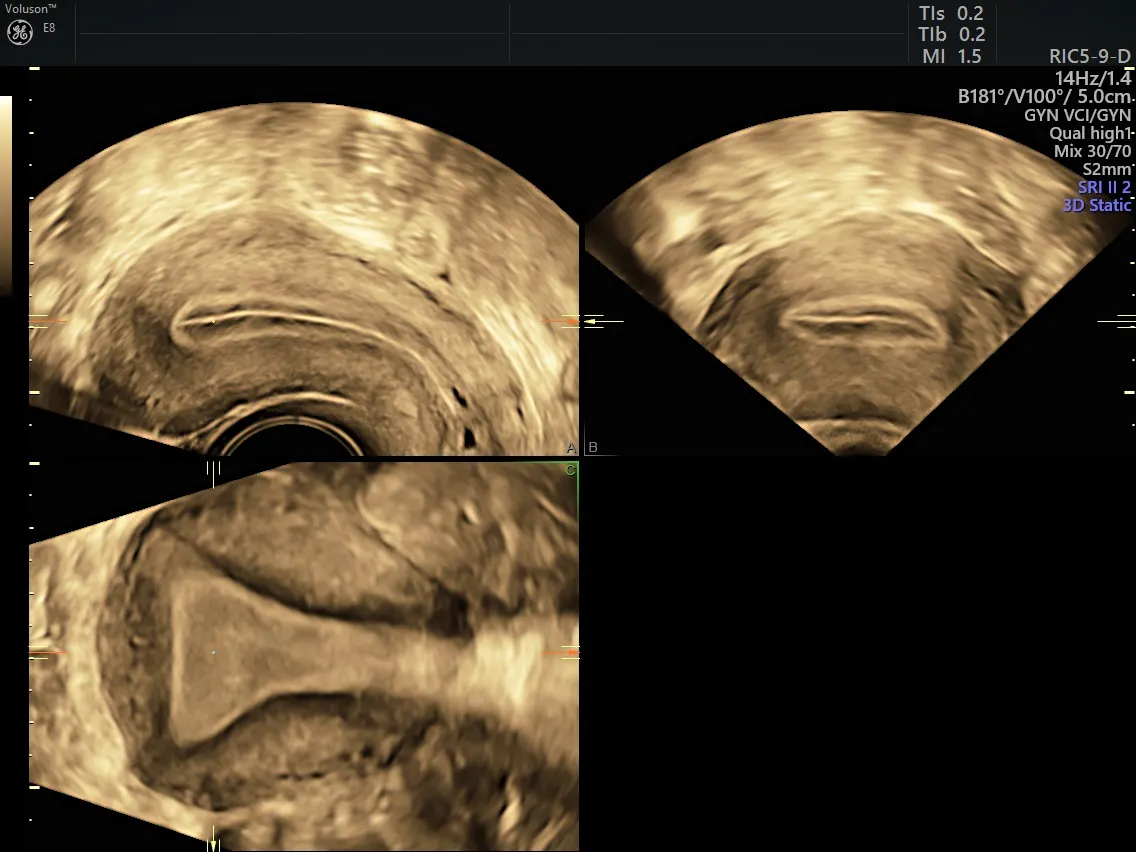
3D Ultrasound - Normal Endometrial Thickness
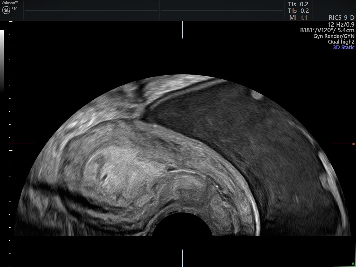
Ultrasound Thickened Endometrium
Using Transvaginal Ultrasound to Predict Endometrial Malignancy
According to the Middle East Fertility Society Journal, the top five ultrasound variables for predicting endometrial malignancy are endometrial thickness greater than or equal to 12 mm, heterogeneous endometrium, irregular endometrial midline, ill-defined endometrium-myometrium interface and a Doppler Score greater than 2.
The sensitivity of color Doppler ultrasound has significantly improved over the years, allowing for ultrasound settings such as HD-Flow to provide a comprehensive, in vivo, noninvasive assessment of gynecological tumors. This is important because the presence of any color flow indicates the formation of blood vessels and introduces concern for endometrial cancer tumor spread.
Transvaginal ultrasound can also assess for the presence of benign masses that can cause postmenopausal bleeding, such as endometrial polyps and submucosal fibroids. Polyps, frequently seen in patients receiving tamoxifen, appear as echogenic, smooth, intracavitary masses that can be outlined by fluid and have a distinct flow stalk. Submucosal fibroids are seen as hypoechoic solid masses that can be either heterogenous or hyperechoic, depending on the degree of degeneration and calcification.
Determining Next Steps
Transvaginal ultrasound provides a valuable initial assessment for postmenopausal women with bleeding. The process of evaluating and diagnosing endometrial cancer can be stressful, so a fast and reliable assessment method is key to providing patients with immediate results so you can help them determine the next steps.
Using ultrasound for a rapid method of assessment of the endometrial thickness, uterine size and possible spread of EC is crucial to a positive patient experience. If the endometrium is thin, the patient can be reassured of the absence of EC. If it is thick and irregular, she can be immediately scheduled for a CT or MRI of the abdomen and pelvis to determine the extent of the tumor spread and begin counseling on treatment options. When determining the symptoms, diagnoses and causes of endometrial cancer, the use of ultrasound is vital.