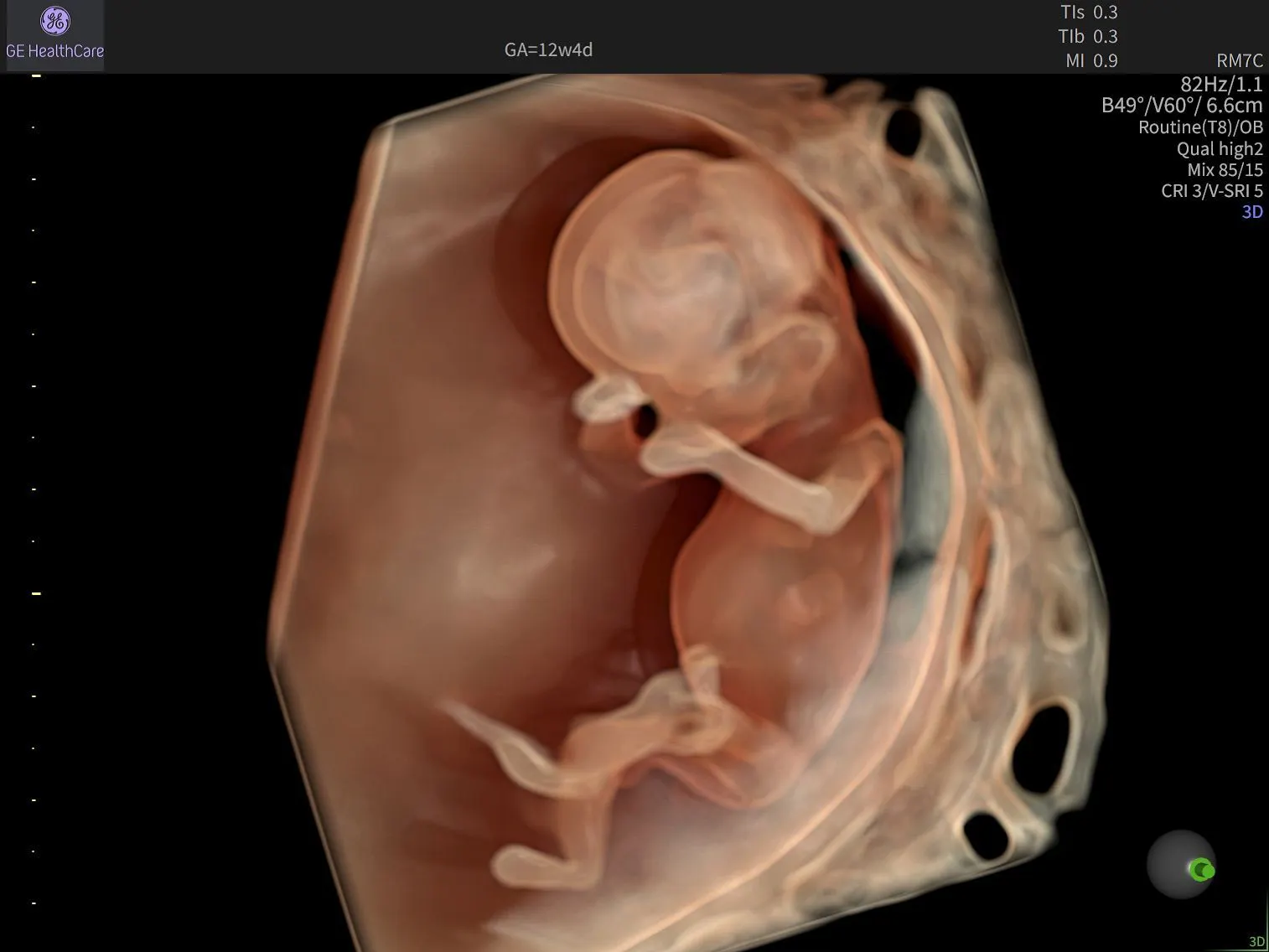
First Trimester Earlier Diagnostics: Unlocking the Potential of Early Detection
The 11–14-week gestational window represents an important opportunity to gain early clinical insights and support informed patient care. During this stage, clinicians can assess fetal anatomy, measure nuchal translucency, evaluate markers associated with chromosomal conditions, and identify structural abnormalities—weeks before the routine anatomy scan at 18–20 weeks.
As ultrasound technology and clinical expertise continue to advance, clinicians are increasingly able to unlock the diagnostic potential of first-trimester imaging. Maximising this opportunity relies on two critical factors: image quality and clinical expertise.¹ Together, they can help transform what is possible during early pregnancy assessment and support greater diagnostic confidence.
The Clinical Value of Earlier Detection
Beyond confirming gestational age and fetal number, the 11–14-week examination provides an opportunity for experienced clinicians to assess fetal development and identify many major structural abnormalities, including central nervous system defects, abdominal wall anomalies, and skeletal abnormalities. Clinicians can also evaluate chromosomal risk markers through nuchal translucency measurements, nasal bone assessment, and ductus venosus flow.
Earlier assessment can provide valuable time for confirmatory testing, specialist consultation, care planning, and patient counselling. Identifying potential concerns at 12 weeks rather than 19 weeks may also allow clinicians and patients to follow findings over a longer period, supporting more informed clinical decision-making throughout pregnancy.
The Importance of Image Quality in the First Trimester
Visualising fetal anatomy between 11 and 14 weeks can present unique challenges. The fetus is small, rapidly developing, and often positioned in ways that can make image acquisition more complex.
Historically, clinicians have balanced a trade-off between penetration and resolution. High-frequency probes deliver detailed images but traditionally have been limited in penetration, while lower-frequency probes penetrate deeper but may compromise image detail.
Additional factors—including probe positioning, patient habitus, precise gestational dating, and operator experience—can also influence image acquisition and diagnostic confidence.²
Because of these factors, exceptional image quality plays a critical role in enabling confident assessment during this early stage of pregnancy. Clear visualisation helps clinicians evaluate anatomy more effectively, identify findings earlier, and provide timely guidance to patients when additional follow-up may be needed.
The Advantages of High-Frequency Endocavitary Imaging
For many first-trimester examinations, transvaginal imaging offers important diagnostic advantages over transabdominal approaches. By positioning the transducer closer to the gestational sac and developing fetal anatomy, the distance ultrasound waves must travel is reduced, minimising interference from surrounding structures.
This approach enables the use of higher-frequency transducers capable of delivering exceptional spatial resolution and anatomical detail. The benefits can be particularly valuable in patients with elevated BMI or a retroverted uterus, where transabdominal imaging may be more challenging.
Research has also demonstrated that transvaginal imaging can provide higher-resolution assessment and may reduce misdiagnosis when evaluating suspected fetal cardiac abnormalities.³ Rather than being viewed solely as a secondary approach, transvaginal imaging can be an important component of a comprehensive first-trimester examination strategy.
Purpose-Built for First Trimester Assessment
The GE HealthCare’s RIC12 endocavitary probe was designed to support the unique demands of first-trimester obstetric imaging.
By combining high-frequency performance, optimised penetration, and a wide field of view, the RIC12 helps clinicians visualise fine fetal anatomy while maintaining exceptional image quality. Rather than requiring a choice between depth and detail, the probe is designed to support both.
These capabilities are particularly important when assessing developing fetal anatomy and performing precise measurements, such as nuchal translucency assessment. Accurate evaluation depends on clear tissue definition and well-defined anatomical boundaries—factors that contribute to confident clinical assessment.
Technology alone, however, is only part of the equation. The value of advanced imaging is realised through the expertise of the clinician interpreting the images. Understanding normal fetal development, recognising subtle anatomical variations, and integrating findings into the broader clinical picture remain essential components of high-quality patient care.
Building Clinical Confidence Through Education
As first-trimester imaging capabilities continue to evolve, ongoing clinician education becomes increasingly important.
Many clinicians develop expertise in early pregnancy assessment within training frameworks traditionally focused on second-trimester anatomy evaluation. As a result, opportunities exist to further strengthen knowledge around first-trimester anatomy, image acquisition, measurement techniques, and clinical interpretation.
Standardised guidance, including recommendations from the International Society of Ultrasound in Obstetrics and Gynecology (ISUOG), provides a valuable framework for consistent acquisition, measurement, and documentation practices.⁴
Maximising the Potential of First-Trimester Ultrasound
The first trimester offers a powerful opportunity to support earlier assessment, informed decision-making, and personalised patient care.
As ultrasound technology advances and clinical expertise continues to grow, clinicians are increasingly able to leverage the diagnostic potential of the 11–14-week examination. High-frequency endocavitary imaging, including the RIC12 probe, combined with ongoing education and evidence-based practice, can help clinicians approach even challenging examinations with greater confidence.
For clinicians looking to expand their expertise, the Empowered Women's Health programme provides educational resources and learning pathways designed to support continuous growth, strengthen clinical confidence, and help unlock the full value of first-trimester ultrasound.
Interested in learning more, watch lectures from:
R.S. Abu-Rustum – First trimester detailed ultrasound in practice
E. Sinkovskaya – Guidelines based approach to first trimester ultrasound
A. Papageorghiou & S. Meagher – The first trimester fetal exam in numbers and images
S. Meagher – Cardiac diagnosis in the first trimester
S. Meagher – Early pregnancy and first trimester malformations
References:
1.Syngelaki A, Chelemen T, Dagklis T, Allan L, Nicolaides KH. Challenges in the diagnosis of fetal non-chromosomal abnormalities at 11-13 weeks. Prenat Diagn. 2011;31(1):90-102. doi:10.1002/pd.2642
2.ISUOG. ISUOG Practice Guidelines: performance of first-trimester fetal ultrasound scan. Ultrasound Obstet Gynecol. 2013;41(1):102-113. doi:10.1002/uog.12342
3.Ji X, Xia Y, Zhang H, Wang H, Shi R, Han Z. Study on the application of ultrasonography in the diagnosis of fetal cardiac structural abnormalities and the relationship between fetal cardiac structural abnormalities with chromosome abnormalities in early pregnancy. Ann Transl Med. 2021;9(24):1790. doi:10.21037/atm-21-6492
4.Bilardo CM, Chaoui R, Hyett JA, et al. ISUOG Practice Guidelines (updated): performance of 11–14‐week ultrasound scan. Ultrasound Obstet Gynecol. 2023;61(1):127-143. doi:10.1002/uog.26106

