
For expecting parents, seeing their unborn baby's heartbeat is an important milestone in the growth of their family. For physicians with a patient at risk for fetal heart abnormalities, this moment is critical for another reason — it's the first opportunity to evaluate the fetal heart for congenital defects.
Thanks to advances in ultrasound technology and fetal diagnostic testing, planning the course of treatment for fetal heart abnormalities can now begin at earlier stages. An early fetal cardiac examination that reveals anomalies sets plans in motion for the remainder of the gestation. Exceptional yet compassionate clinical care across all points of treatment strengthens the doctor-patient relationship and makes for a more positive experience for patients.
What Are Indicators of Risk for Fetal Heart Anomalies?
Deciding whether early fetal heart ultrasound is necessary depends on several factors, ranging from genetics to the type of pregnancy. The genetic indicators of risk are family history, chromosomal abnormalities and single gene defects. Assisted reproductive technology pregnancies and monochorionic twin pregnancies also increase suspicion for cardiac anomalies.
Certain abnormal findings seen on first-trimester ultrasound should alert physicians to take a closer look at the overall risk for heart issues. These include structural abnormalities, an abnormal heart-to-chest area ratio and an enlarged nuchal translucency.
Deciding if early fetal cardiac ultrasound is necessary is more straightforward when used in combination with noninvasive prenatal testing (NIPT). NIPT provides adequate results concerning congenital heart defects (CHD) that result from typical aneuploidies. But NIPT does have limitations given that it can only assess risk levels — not diagnose conditions. Therefore, a low-risk test result cannot rule out all types of heart defects. The diagnosis of these other defects requires invasive fetal testing. Findings of an enlarged nuchal translucency measurement on first-trimester ultrasound can help parents considering NIPT versus invasive fetal testing make an informed decision.
What Is an Early Fetal Cardiac Examination, and Who Needs One?
An early fetal cardiac ultrasound can check for heart abnormalities between the 11th to 19th week of gestation. Advances in ultrasound technology, including spatiotemporal image correlation volumes, are continuing to improve the accuracy of diagnosis at this very early stage. Before these advances came into existence, the four-chamber view was the standard for imaging the fetal heart. Now, better resolution of outflow tract views and the three-vessel trachea view provide more information even at this early stage. Using state-of-the-art ultrasound systems enables physicians to add additional imaging views as visualization improves closer to 13 weeks.
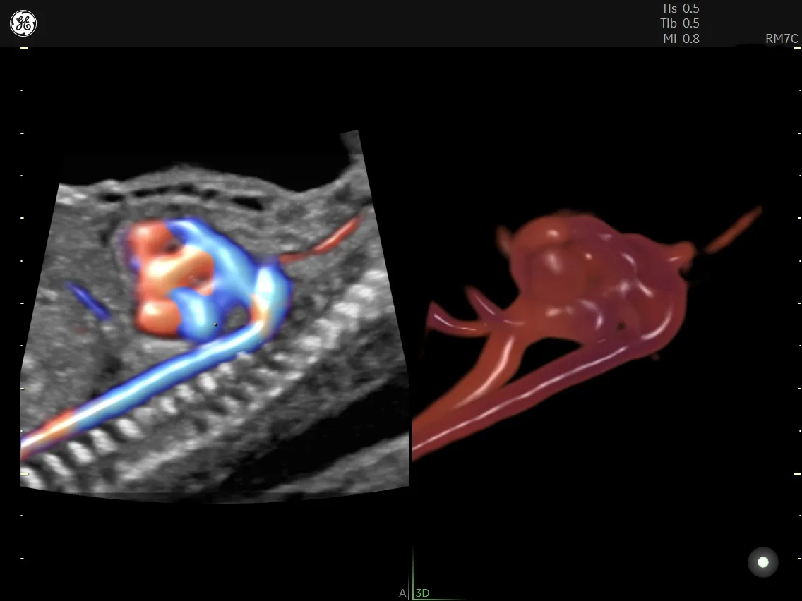
Image
Voluson™ HDlive™ Flow Silhouette – Visualize blood vessels and fetal heart flow to provide greater insight transparently through vascular anatomy.
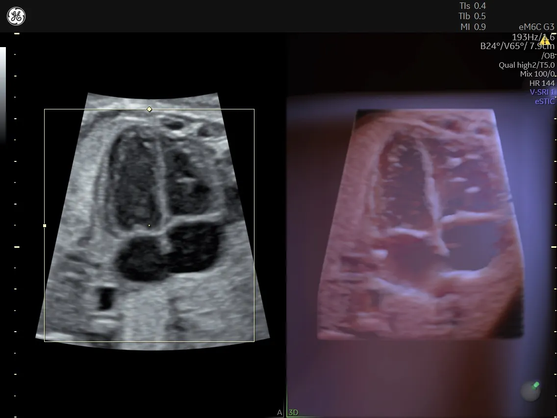
eSTIC
eSTIC with HDlive™ Studio+, Enhance fetal cardiac exams with improved resolution.
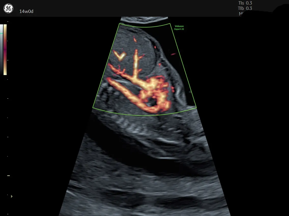
RIC6-12
Voluson's RIC6-12 high-resolution endovaginal probe helps detect fetal abnormalities earlier in the first trimester.
The early fetal cardiac ultrasound is useful in a variety of cases. According to the Center for Fetal Therapy at Johns Hopkins Medicine, an abnormal combined first-trimester nuchal translucency screening is an indication for the exam. The atypical findings on this exam include a nuchal translucency greater than 3.5 millimeters, an abnormal cardiac axis orientation, an abnormal fetal heart rate and reversal of flow in both the ductus arteriosus and tricuspid valve.
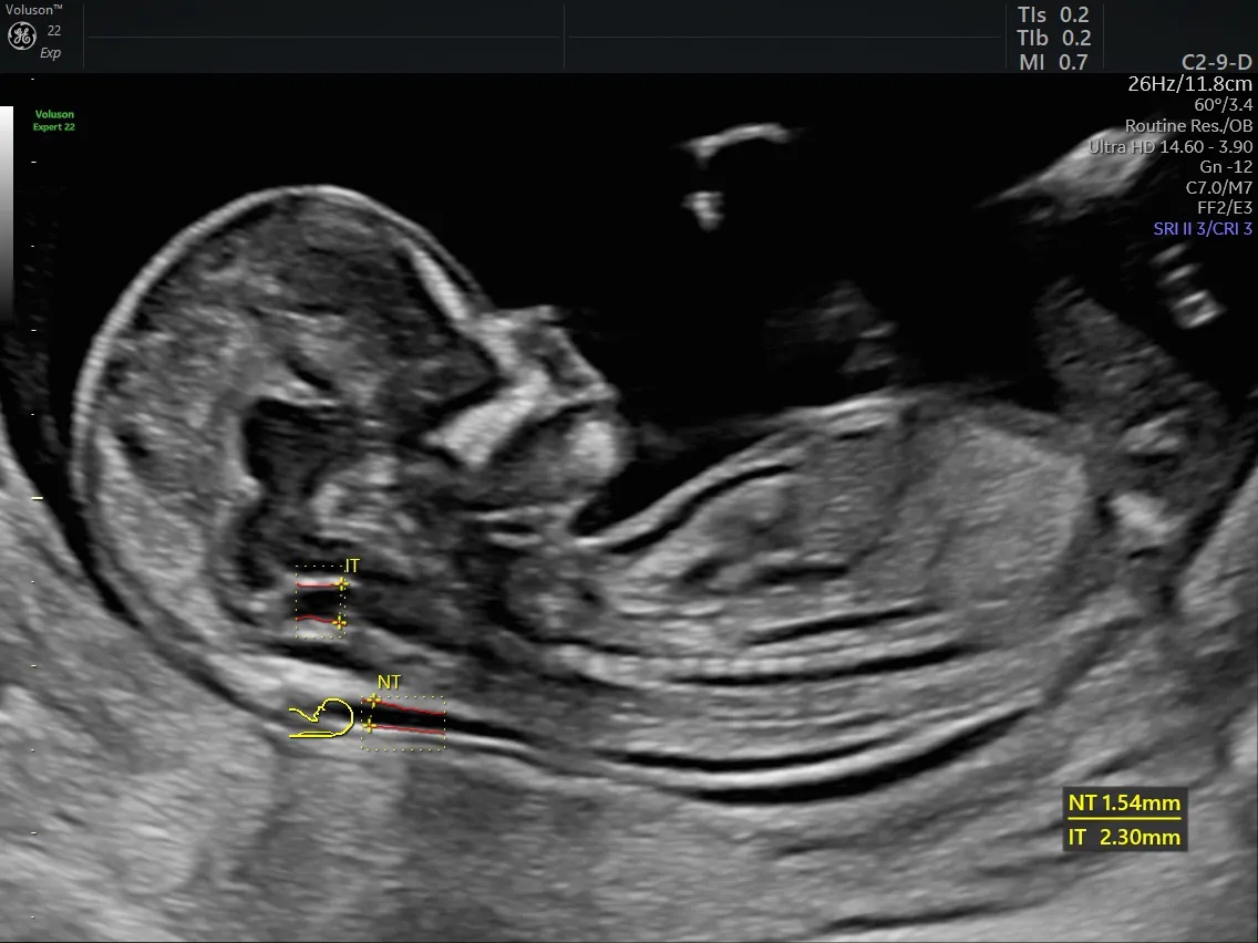
Fetus
Consistently assess the nuchal and intracranial translucency with Voluson SonoNT and SonoIT automation.
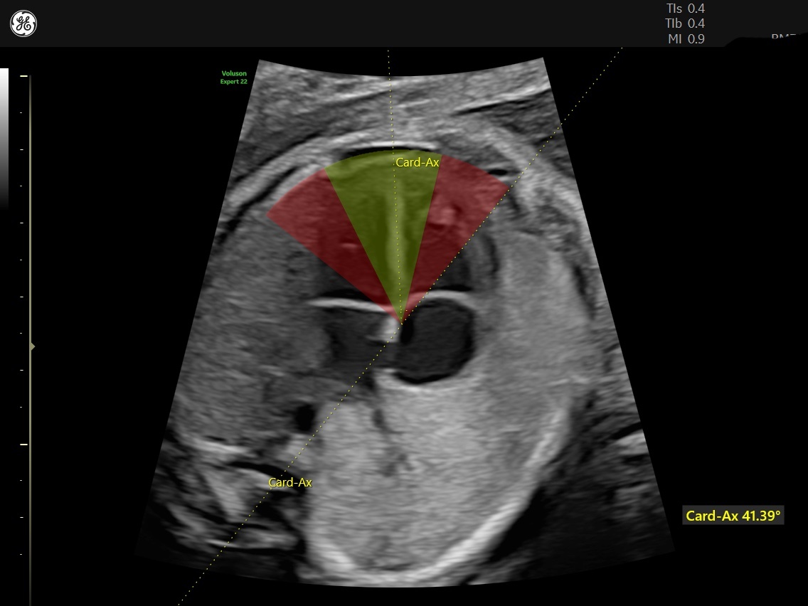
Voulson Cardiac Axis
Voluson's Cardiac Axis is an automated measurement to help quickly identify the potential for a congenital heart defect.
Several maternal conditions may also put a fetus at a higher risk for fetal heart anomalies, according to research published in Circulation. Patients with a personal or family history of CHD, or with diseases including lupus, phenylketonuria and pre-existing maternal diabetes can all benefit from an early fetal cardiac exam. The use of anti-seizure medications, lithium and other teratogens can also pose the question of potential heart defects, making patients using these substances candidates for the test as well.
Can Early Detection Decrease the Risk of Life-Threatening Complications?
There are several advantages to knowing about fetal cardiac conditions well ahead of delivery.
The first-trimester ultrasound exam provides a significant advantage by alerting the physician to refer the patient to a high-risk specialist for ultrasound evaluation of any anomalies, which could otherwise be missed by a less detailed Level 1 OB scan.
Another advantage of early detection is early access to genetic counseling. This counselling is critical to assessing personal and family history that may add to risk for the fetus. It also provides the education and support parents need to make prenatal diagnostic testing decisions.
But early detection of fetal heart anomalies does more than indicate the need for advanced fetal testing and therapies — it calls for prenatal counseling for parents. Having a trained professional to talk to about concerns can even help reduce the effects of the parent's stress and anxiety on the fetus, according to a study published in Obstetric Medicine. Given that prenatal maternal stress can affect a child throughout their lifespan, offering this assistance is a priority. Conditions that may develop in the offspring of those under stress during their pregnancies include asthma, allergies, attachment difficulties, mood disorders, hypersensitivity to stress and even poor immune response.
Why Should Physicians Plan Multidisciplinary Prenatal and Perinatal Care?
Once a fetal heart issue is detected, the physician can begin to put together a treatment plan and care team. Appointing a team not only sets the stage for the best possible outcomes, intervention and treatment, but it also can be a great comfort to anxious families. Deciding on the hospital or medical center where the delivery will (ideally) occur is a key first step. It ensures the necessary medical support or transportation for the newborn is immediately available. Simply knowing where the parent will give birth can offer them peace of mind.
The prenatal care team for a parent whose fetus has a heart condition usually consists of their OB/GYN, maternal-fetal specialists, genetic counselors, neonatologists and pediatric cardiologists. In cases where surgery will be necessary, pediatric cardiovascular surgeons will also be in on the consultation so all possible plans are ready to implement shortly after birth.
Many of these team members continue providing care after delivery. A vital addition to the team are the nurses who will care for the newborn. Infants needing specialized care may need to be taken to the neonatal intensive care unit directly following birth. Here, supervision and treatment will continue until the neonatologist feels the baby is stable enough to go home.
How Can Physicians Improve Treatment and Outcomes for Fetal Heart Abnormalities?
The Indian state of Kerala has recently become a model for how to plan treatment for fetal cardiac abnormalities. Upon investigating the region's high mortality rates in the first 28 days after birth, the local health department found one of the main culprits to be congenital heart disease. The lack of treatment centers offering pediatric heart surgery and the challenge of transporting patients added to the problem. Dr. Balu Vaidyanathan, a clinical professor of pediatric cardiology, and Dr. B. Ekbal, a member of the Kerala State Planning Board, stepped in with a plan.
"Instead of waiting for the baby to be born and then finding out if the baby has a heart problem, we could do a heart examination in the anomaly scan," Dr. Vaidyanathan told The News Minute. "This also means that the treatment of the baby could start soon after delivery, without any delay."
This change led to an improvement in care and a reduction in costs, Dr. Vaidyanathan reported.
Dr. Vaidyanathan was able to train more than 500 physicians on how to detect fetal heart anomalies during ultrasound scans, partly by supplying flashcards depicting an imaging protocol of only the essential views necessary for diagnosis. Along with hands-on training for doctors, these interventions made it possible to decrease Kerala's neonatal mortality rate from 12 deaths per 1,000 live births in 2002 to only seven deaths per 1,000 live births in 2020.
Does Early Diagnosis Make a Difference?
Diagnosing fetal heart abnormalities early can significantly impact the care provided. By utilizing cutting-edge technology to view the fetal heart's tiny structures, physicians can achieve reliable detail and greater confidence in ultrasound findings — both of which will continue to increase as users gain experience. From scheduling serial imaging and further diagnostics to managing infant care at birth, making these views part of a protocol substantially improves patient outcomes.

