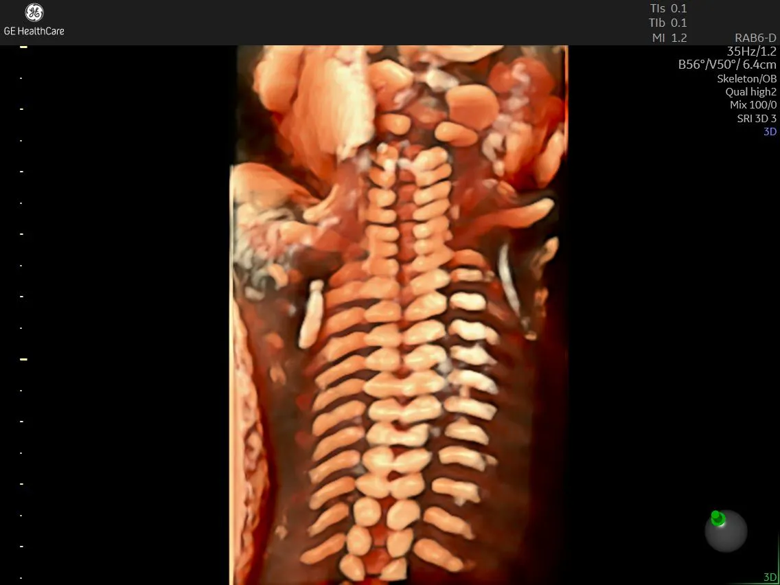
How HDlive Silhouette Is Crucial in Evaluating the Fetal Spine in Myelomeningocele
Neural tube defects, a constellation of congenital malformations of the fetal spine, affect nearly 140,000 patients worldwide each year, according to a 2023 article in StatPearls. Open spina bifida is the most common congenital fetal central nervous system anomaly. In its most severe form, myelomeningocele, the spinal cord and membranes protrude from the back of the affected fetus, with risks that range from pregnancy complications to lifelong disability.
Early detection aids in prompt prenatal surgical management, yet careful evaluation of the fetal central nervous system with 2D ultrasound requires a significant degree of imaging expertise, which may not be readily available in many ultrasound facilities, notes the International Society of Ultrasound in Obstetrics and Gynecology (ISUOG). Now, new 3D ultrasound promises greater ease, visibility, and consistency when assessing spine health.
Ultrasound and Fetal Spine Examination
Direct examination of the fetal spine with ultrasound is the foremost method for diagnosing spina bifida, reports the most recent practice guidelines from ISUOG. Similarly, in cases where the lesion has advanced to myelomeningocele, ultrasound plays an essential role in defining the upper anatomical level of the growth—one of the chief predictors of neurological morbidity after delivery.
Ultrasound also reveals the health of the fetal vertebrae. Using longitudinal, coronal, and transverse views, sonographers can look for and assess the characteristic V-shape, which is formed by a widening of the transverse processes in concert with dorsal defects. Accurately characterizing the content and size of the defect allows for a thorough assessment of potential postnatal delays and disability while also aiding surgical planning. Yet, in some cases, the position of the fetus or operator inexperience may make it challenging to inspect all the necessary structures with traditional 2D ultrasound and thus arrive at an accurate diagnosis.
What Is HDlive Silhouette?
HDlive™ Silhouette removes the barriers to accurate diagnosis, offering a clear view of the level of anatomical defect and the span of the lesion in fetuses with severe malformation like myelomeningocele. HDlive Silhouette is the newest generation of HDlive, a 3D rendering method that produces realistic fetal images. With HDlive Silhouette, sonographers can easily evaluate solid surface structures and the internal anatomy of the fetus because the technology dynamically applies transparency to specific structures within the rendered volume. By taking advantage of advanced beamforming technology, speckle reduction algorithms, and composite resolution imaging technologies, HDlive provides exceptional image quality for even the most challenging clinical examinations.
HDlive Silhouette in Practice
A recent case involving a patient in the 24th week of pregnancy illustrated the use of the 3D technology in practice. The examination began with a transabdominal and vaginal scan of the fetal spine with two different probes: a matrix convex volumetric RM6C probe and a convex volumetric RIC6-12-D probe. The sonographer positioned the probes with the transducer near the center of the fetus's back, aligned to a midsagittal plane.
When correctly aligned, the system renders an image of the vertebral bodies, spinous processes, and the condition of the skin covering the spine. Notably, myelomeningocele is a congenital anomaly characterized by a skinless area, where the spinal cord is pushing out, with a cyst that sometimes contains cerebrospinal fluid and neural tissue.
In this case, after identifying the spinal defect, the sonographer activated the 3D button on the platform. The region of interest (ROI) was then adjusted to capture the defect and the whole vertebral column longitudinally, with enough depth to show the vertebral bodies. The sweep angle was set to 35-40 degrees for each side, and high-quality degrees were established to capture a surface rendering.
The ROI was then tightened to fit only the defect, allowing the sonographer to visualize the dorsal cyst. Reducing the gains and depth revealed the vertebral column and its anatomical references. These landmarks included the most caudal articulated rib (T12), the superior aspect of the iliac crest (L5), and the lowest and most ossified vertebra representing S4. From here, the sonographer noted the unaffected vertebral arches and counted them, starting from T12 and moving down the spine toward the tailbone. Doing so clearly showed where the lateral processes diverged. In this case, the sagittal view showed a divergence beginning at L2 of the posterior arches of the spine.
Thus, HDlive Silhouette played a significant role in accurately visualizing some structures that weren't easily visualized in 2D ultrasound, promoting diagnostic accuracy and informing medical management for enhanced patient care.
Learn more here: HDlive Silhouette in evaluation of fetal spine in myelomeningocele.

