How Intraoperative Ultrasound Supports Confident Decision-Making in Gynecologic Surgery
Every gynecologic procedure relies on a thorough understanding of anatomy and pathology. While preoperative ultrasound and MRI play a critical role in surgical planning, the intraoperative environment can present unexpected findings that influence procedural decisions.1
Intraoperative ultrasound (IOUS) extends the value of imaging into the operating room, providing real-time visualisation that complements preoperative assessment. By enabling surgeons to localise pathology, clarify anatomy, and adapt their approach during surgery, IOUS can help support informed clinical decision-making in a range of gynecologic procedures.1,4
What Is Intraoperative Ultrasound?
Intraoperative ultrasound (IOUS) refers to the use of ultrasound imaging during an active surgical procedure to provide real-time anatomical guidance.
Ultrasound is particularly well suited for intraoperative imaging because it is portable, delivers immediate visual feedback, and does not use ionising radiation. By positioning the transducer directly on or near pelvic structures, clinicians can obtain high-resolution images that support assessment throughout the procedure.1
Depending on the surgical approach, IOUS may be performed using transabdominal, transvaginal, or laparoscopic techniques.
Transabdominal IOUS
Typically used during open or laparoscopic procedures, transabdominal IOUS can visualise uterine and adnexal structures through the abdominal wall or directly on organ surfaces.
Transvaginal IOUS
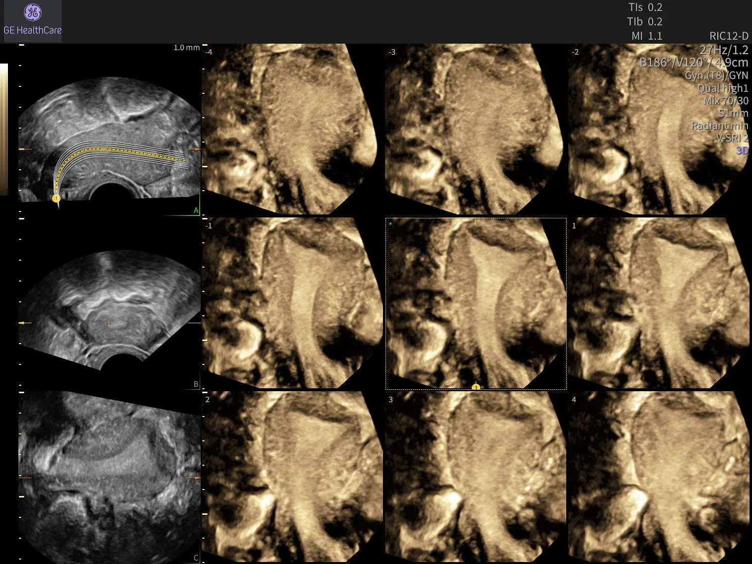
Transvaginal imaging is particularly valuable during hysteroscopic and uterine procedures, providing high-resolution visualisation of the endometrial cavity and surrounding myometrium.
Laparoscopic Ultrasound (LUS)
Laparoscopic ultrasound involves introducing a dedicated ultrasound probe through a trocar port, allowing direct imaging of pelvic organs during minimally invasive surgery.
Why Real-Time Imaging Matters in the Operating Room
Surgical findings do not always mirror preoperative imaging. Adhesions, large fibroids, distorted anatomy, or extensive endometriosis can alter the appearance of tissues once a procedure begins.1,7
By providing real-time imaging during surgery, IOUS can help surgeons better understand anatomy and pathology as operative findings evolve.1,4 As with all advanced ultrasound applications, success depends on the clinician's ability to accurately acquire, interpret, and act upon imaging findings in the moment.1,4
Supporting Real-Time Clinical Decisions
Intraoperative ultrasound can help localise pathology, assess anatomical relationships, and guide the extent of surgical intervention.1
Having access to dynamic imaging during a procedure may support decision-making when anatomy is complex or findings differ from preoperative expectations.1,4
Enhancing Surgical Precision
Real-time visualisation can assist surgeons in performing more targeted dissections and resections.
For example, during laparoscopic myomectomy, IOUS has been shown to improve localisation of fibroids that may be difficult to identify visually, particularly when lesions are small, deeply embedded within the myometrium, or located beneath the uterine surface.2
By helping identify pathology more precisely, ultrasound can contribute to a more informed surgical approach.
Supporting Patient Safety
Understanding the spatial relationship between pathology and adjacent structures is fundamental to surgical planning and execution.1,7
In hysteroscopic procedures, IOUS can help clinicians monitor resection depth and evaluate residual myometrial thickness throughout the procedure. This additional visualisation may help support precise tissue removal while reducing the risk of uterine perforation.3,7
Clinical Expertise Remains Central
Like many advanced ultrasound applications, IOUS is highly operator dependent. Its value depends not only on the technology itself but also on the clinician's expertise in image acquisition, anatomical knowledge, and procedural technique.1,4,7
Successful integration of intraoperative ultrasound requires training, experience, and confidence in real-time image interpretation.4 Clinicians and surgical teams should develop proficiency in both acquiring and interpreting images before incorporating IOUS into routine practice.
Clinical Applications of IOUS in Gynecologic Surgery
Evidence supporting IOUS varies across clinical applications, but several areas of gynecologic surgery have demonstrated meaningful practical value.
Myomectomy
During myomectomy, IOUS can assist with localisation of intramural and submucosal fibroids that may not be visible on the uterine surface. Real-time imaging can support incision planning and help reduce the likelihood of missed lesions.2
Hysteroscopic Procedures
IOUS can guide the resection of submucosal fibroids, endometrial polyps, adhesions, and uterine septa by helping clinicians evaluate resection depth and residual myometrial thickness throughout the procedure.3 This may support more precise intervention while reducing the risk of uterine perforation.3,7
Endometriosis Excision
In cases of deep infiltrating endometriosis, IOUS may help surgeons evaluate lesion depth, disease extent, and relationships to surrounding structures during complex excision procedures.4,6
Gynecologic Oncology
In gynecologic oncology, IOUS is being explored as a tool to support tumour localisation, assessment of suspicious tissue, fertility-sparing procedures, cytoreductive surgery, and emerging contrast-enhanced imaging applications.5,7
Additional Applications
IOUS may also support ultrasound-guided removal of malpositioned intrauterine devices (IUDs) and management of retained products of conception in anatomically challenging cases.7
Considerations for Implementation
Successful implementation begins with selecting the appropriate ultrasound system and transducer for the planned procedure while maintaining sterile operating room technique.1
Because IOUS is operator dependent, clinicians benefit from structured training, supervised clinical experience, and ongoing practice in image acquisition and interpretation.1,4
Careful workflow planning can also help support efficient adoption. Considerations include:
- Probe positioning
- Sterile field management
- Documentation processes
- Communication with surgical team members
When intraoperative ultrasound influences clinical management, both its role and key findings should be documented within the operative report.1
The Evolving Role of Intraoperative Ultrasound
Growing evidence supports the use of IOUS across a range of gynecologic procedures, with the strongest evidence currently available for myomectomy and hysteroscopic surgery. Applications in endometriosis excision and gynecologic oncology continue to generate interest, although these areas remain less established.2,3,4,5
Many published studies are based on narrative reviews, smaller clinical investigations, and procedure-specific experiences rather than large randomised trials. Nevertheless, the ability to visualise anatomy and pathology in real time offers a compelling opportunity to support surgical decision-making when integrated appropriately into clinical practice.
As ultrasound technology continues to advance, the greatest value will continue to come from the expertise of the clinician using it. Ongoing education, hands-on experience, and a commitment to continuous learning help build the confidence needed to effectively incorporate IOUS into the surgical workflow.
Bringing Imaging into the Moment of Decision
Intraoperative ultrasound provides more than real-time imaging. It offers clinicians an additional layer of insight at the point when decisions matter most.
When supported by appropriate training and experience, IOUS can help surgeons visualise anatomy with greater clarity, adapt to unexpected findings, and make more informed intraoperative decisions. By bringing imaging directly into the operating room, ultrasound continues to expand its role as a valuable partner in precision gynecologic surgery.
References
1.Lubner MG, et al. Diagnostic and procedural intraoperative ultrasound: technique, tips and tricks for optimizing results. Br J Radiol. 2021;94(1121):20201406. doi:10.1259/bjr.20201406
2.Aktoz F, Arslan T, Güzel Y. Intraoperative laparoscopic ultrasound during laparoscopic myomectomy: a narrative review. J Turk Ger Gynecol Assoc. 2024;25(3):179-183.
3.Ferreira de Castro L, Santos I, Laganà AS, de Vree B, et al. Enhancing precision in hysteroscopic surgery: the role of intraoperative ultrasound. Eur J Obstet Gynecol Reprod Biol. 2024;302:306-309. doi:10.1016/j.ejogrb.2024.09.027
4.Grewal K, Jones B, L'Heveder A, et al. The use of intra-operative ultrasound in gynecological surgery: a review. Future Sci OA. 2021;7(3):FSO678. doi:10.2144/fsoa-2020-0172
5.Lakany M, Sharif A, et al. Intraoperative ultrasound as a decision-making tool in modern gynecologic oncology. J Pers Med. 2025;15(7):296. doi:10.3390/jpm15070296
6.Guerriero S, et al. Laparoscopic ultrasound for intraoperative mapping of deep infiltrating endometriosis. Ultrasound Obstet Gynecol. 2018;52(3):378-385.
7.Kasaven LS, Galazis N, et al. Intraoperative gynecological ultrasound. Glob Libr Women's Med. Published online. doi:10.3843/GLOWM.419763

