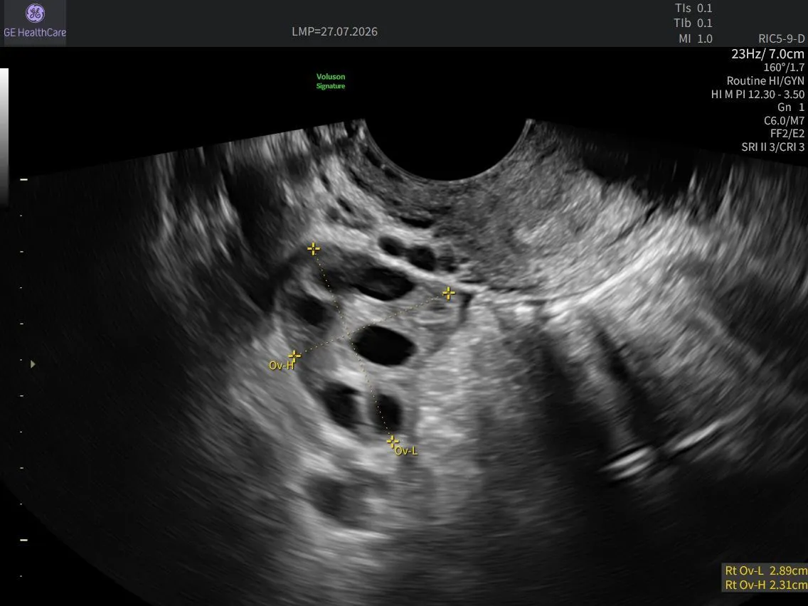
Routine GYN Exams: Enhancing Efficiency While Supporting Measurement Consistency
Routine gynecologic ultrasound examinations are among the most commonly performed imaging studies in women's healthcare. Yet behind every routine scan are measurements that can influence clinical decisions, support longitudinal patient management, and provide critical insight into reproductive health.
Uterine dimensions, endometrial thickness, and ovarian volume are key measurements used to assess structural abnormalities, evaluate cancer risk, and support the diagnosis and monitoring of conditions such as polycystic ovary syndrome (PCOS). Accurate and reproducible measurements are essential, as even small variations can impact clinical interpretation and patient management.
As healthcare systems face increasing patient volumes and growing demands on staff, clinicians must balance efficiency with consistency. Intelligent automation offers an opportunity to streamline repetitive tasks while preserving diagnostic confidence. Rather than replacing clinical expertise, automated biometry helps clinicians focus on what matters most: image interpretation, clinical decision-making, and patient care.
The Clinical Value Behind Routine Measurements
A standard transvaginal gynecologic ultrasound examination includes several important measurements:
- Uterine length, width, and anteroposterior diameter
- Endometrial thickness measured at its thickest point in the sagittal plane
- Ovarian dimensions and volume, typically calculated using the prolate ellipsoid formula
These measurements provide valuable clinical information throughout a woman's life.
Uterine dimensions offer insight into reproductive health and hormonal status. Endometrial thickness plays an important role in fertility assessment, evaluation of abnormal uterine bleeding, and screening for conditions such as endometrial hyperplasia. Ovarian size and volume can support assessment of conditions including PCOS and diminished ovarian reserve.
Average uterine dimensions are approximately 8 cm in length, 5 cm in width, and 4 cm in thickness, although size varies according to age, parity, and menopausal status.¹ Likewise, ovarian dimensions change throughout life, with normal premenopausal ovaries generally measuring 3 to 5 cm in length and 1.5 to 3 cm in width, with an average volume of 3 to 10 mL.²˒³
Endometrial thickness also varies significantly. In premenopausal women, measurements may range from 1 to 4 mm immediately following menstruation to approximately 14 mm during the secretory phase. In postmenopausal women, normal endometrial thickness is typically less than 5 mm.⁴
Because clinical decisions often rely on threshold values, small differences in measurement can have meaningful implications. A variation of only a few millimetres may influence whether additional imaging, surveillance, or biopsy is recommended.
The Challenge of Manual Measurements
Manual measurement has long been a standard component of ultrasound examinations. However, variations in operator experience, caliper placement, and image optimisation can contribute to differences in measurement results.
Research has shown that operator experience remains a key contributor to variability. In a study comparing manual and semi-automated ultrasound measurements, non-expert operators demonstrated substantially greater variability using manual measurements, while semi-automated caliper placement improved reproducibility to levels similar to those achieved by expert operators.⁵
The authors concluded that automation helped standardise the measurement process while maintaining clinician oversight. At the same time, image acquisition remained the most operator-dependent component of the examination.⁵
In busy clinical environments, factors such as workload, time pressure, and repetitive manual tasks may further contribute to variability. When serial measurements are used to monitor a patient's condition, inconsistency can make it difficult to determine whether observed changes reflect true disease progression or differences in measurement technique.
Bringing Automation into the Routine GYN Exam
Automation tools such as Voluson™ SonoBiometry GYN are designed to support clinicians by reducing repetitive measurement tasks, standardising routine biometry, and helping deliver consistent results across examinations.
Using AI-powered automation, Voluson SonoBiometry GYN automatically identifies anatomical landmarks, places calipers, and captures uterine and ovarian measurements. By reducing manual keystrokes and automating repetitive tasks, clinicians can spend less time documenting measurements and more time focusing on image assessment, clinical interpretation, and patient care.
Rather than manually identifying landmarks and placing measurement calipers, clinicians can leverage automation to perform routine measurement tasks efficiently while maintaining full clinical control throughout the examination.
Importantly, Voluson SonoBiometry GYN integrates seamlessly into existing scanning protocols. Automated measurements are obtained during the routine examination without requiring additional scanning planes, patient repositioning, or changes to standard workflows.
The sonographer remains responsible for image acquisition, while the interpreting clinician reviews and validates automated measurements before they become part of the patient's record. Automation supports the workflow, but clinical expertise remains central to every clinical decision.
The Efficiency Advantage: Less Repetition, More Focus
Ultrasound examinations require continuous observation, analysis, and decision-making. Repeated manual tasks performed throughout the day contribute to both cognitive and physical workload.
Artificial intelligence can help streamline repetitive aspects of ultrasound imaging by automatically identifying anatomical structures and performing measurement-related tasks. By helping reduce manual interaction and supporting standardised measurement techniques, automation can improve workflow efficiency while maintaining consistency.
For clinicians, this means fewer repetitive steps, reduced documentation burden, and more time to focus on image quality, clinical interpretation, and patient interaction.
By reducing keystrokes associated with routine measurements, automation may also help minimise workflow interruptions, supporting a more streamlined scanning experience.
Reproducibility Matters in Longitudinal Care
For patients with conditions requiring ongoing monitoring, measurement consistency is just as important as measurement accuracy.
Conditions such as uterine fibroids, endometrial hyperplasia, and polycystic ovarian morphology are often assessed over time. Reliable longitudinal evaluation depends on the ability to compare measurements confidently across multiple examinations.
When variability results from differences in operator technique rather than changes in pathology, clinicians may risk overestimating or underestimating disease progression. Automated biometry helps address this challenge by applying a consistent measurement approach regardless of who performs the scan.
By supporting greater reproducibility across examinations, automation can help clinicians make more informed decisions regarding disease progression, treatment response, and patient management.
Empowering Expertise Through Intelligent Automation
The future of ultrasound is not about choosing between technology and clinical expertise. It is about bringing them together.
As automation capabilities continue to evolve, clinicians equipped with both advanced imaging tools and expert clinical knowledge are uniquely positioned to deliver confident, consistent patient care.
Automation tools such as Voluson™ SonoBiometry GYN help bridge this gap by combining AI-powered automation with clinician oversight. Automated caliper placement and measurement capture can reduce keystrokes, support measurement consistency, and streamline repetitive workflow tasks, while clinicians remain the final authority for image acquisition, interpretation, and diagnosis.
Through ongoing clinical education and thoughtful adoption of workflow-enhancing technologies, standardised automation and expert interpretation can work together to support reproducible results, efficient workflows, and confident decision-making.
Ultimately, every gynecologic ultrasound exam generates valuable clinical information. By helping streamline workflow, reduce repetitive tasks, and support measurement consistency, Voluson SonoBiometry GYN empowers clinicians to focus on what they do best: delivering informed, confident care to every patient.
References:
1.Ameer MA, Peterson DC. Anatomy, Abdomen and Pelvis: Uterus. StatPearls Publishing; 2026 Jan-. Available from: https://www.ncbi.nlm.nih.gov/books/NBK470297/
2.Kelsey TW, Dodwell SK, Wilkinson AG, et al. Ovarian volume throughout life: a validated normative model. PLoS One. 2013;8(9):e71465. doi:10.1371/journal.pone.0071465
3.Upadhyaya RP, Ansari MA, Jha A, Kayashta P, Paudel S. Sonographic quantification of ovarian volume in adults attending general health checkup and general out patient department in Tribhuvan University Teaching Hospital. Kathmandu Univ Med J. 2020;18(71):275-278. doi:10.3126/kumj.v18i3.49223
4.Sadro CT. Imaging the endometrium: a pictorial essay. Can Assoc Radiol J. 2016;67(3):254-262. doi:10.1016/j.carj.2015.09.012
5.Abele H, Hoopmann M, Wright D, et al. Intra- and interoperator reliability of manual and semi-automated measurement of fetal nuchal translucency by sonographers with different levels of experience. Ultrasound Obstet Gynecol. 2010;36(4):417-422. doi:10.1002/uog.8809

