

Thoroughly assessing breast tissue has long been a daunting and difficult task for both women performing self-exams at home and clinicians interpreting modern diagnostic imaging. Breasts are surprisingly complicated, and despite the best efforts of many, breast cancer often still goes unnoticed. Fortunately, medical professionals have recently started using 3D automated breast ultrasounds for additional breast cancer screening, reducing the amount of missed breast cancer diagnoses and saving more lives.
How Breast Density Affects Assessment
The difficulty of breast exams and assessment is in part due to the fact that breast density varies widely; no two breasts are the same. A denser breast contains more glandular and connective tissue, and according to BreastCancer.org, the cancer rate is 6 times greater in extremely dense breasts than it is in less-dense breasts. Because of these qualities, mammograms have a decreased ability to detect cancer amid such density.
Traditionally, breast density is measured by assessing at the amount of tissue on a mammogram. The Breast Imaging Reporting and Database System (BI-RADS), which reports mammogram findings, also includes information on breast density and classifies breasts into four groups: mostly fatty, scattered areas of density, heterogeneously dense or extremely dense.
Dense breasts — which fall under the heterogeneously dense and extremely dense BI-RADS categories — are actually quite common, as approximately 43 percent of women in the U.S. ages 40 to 74 years old are classified as having these types of dense breasts.
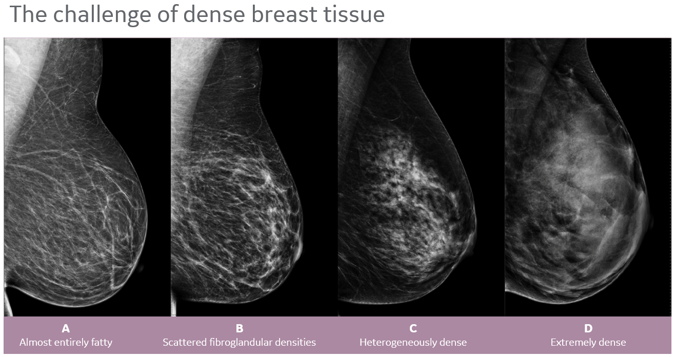
Breast Density Images - BIRADS
How combining 3D Automated Breast Ultrasound and Mammography Improve the Screening Process
Both dense tissue and cancer appear white on a mammogram, so it is like trying to find a snowball in a snowstorm. When ultrasound is used, cancer appears black and dense tissue appears white making cancers easier to see. Since mammography screening is less effective at scanning dense breast tissue, the introduction of supplemental ultrasound imaging would be a logical next step. Adding a 3D automated breast ultrasound for breast cancer screening would be more effective than using mammography alone for this group of women.
When ultrasound technology and mammography are used together as routine breast-screening tools, the combined exams detect more cancers in young women and women with high-density breast tissue, as reported in Science Daily. 3D breast ultrasounds combined with mammography results in correct identification of cancer in more than 90 percent of cases, whereas mammography alone identified just 77 percent of cancers.
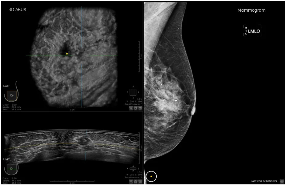
3D ABUS and Mammogram - Biopsy proven IDC
Doctors are also considering the newest form of 3D ultrasound: 3D automated breast ultrasound (3D ABUS). According to the European Journal of Radiology, adding 3D ABUS to mammography screening in dense breasts increases cancer detection without increasing the radiation dose and with an acceptable recall rate. 3D ABUS could become an important screening modality as an addition to mammography, as it is well-suited for screening workflow.
Improving Breast Cancer Treatment and Reducing Deaths
The results of these recent studies suggest that adding 3D ABUS screening to mammography results in more accurate assessments of breast cancer for women with dense breasts, which could ultimately lead to improved treatment of the disease.

