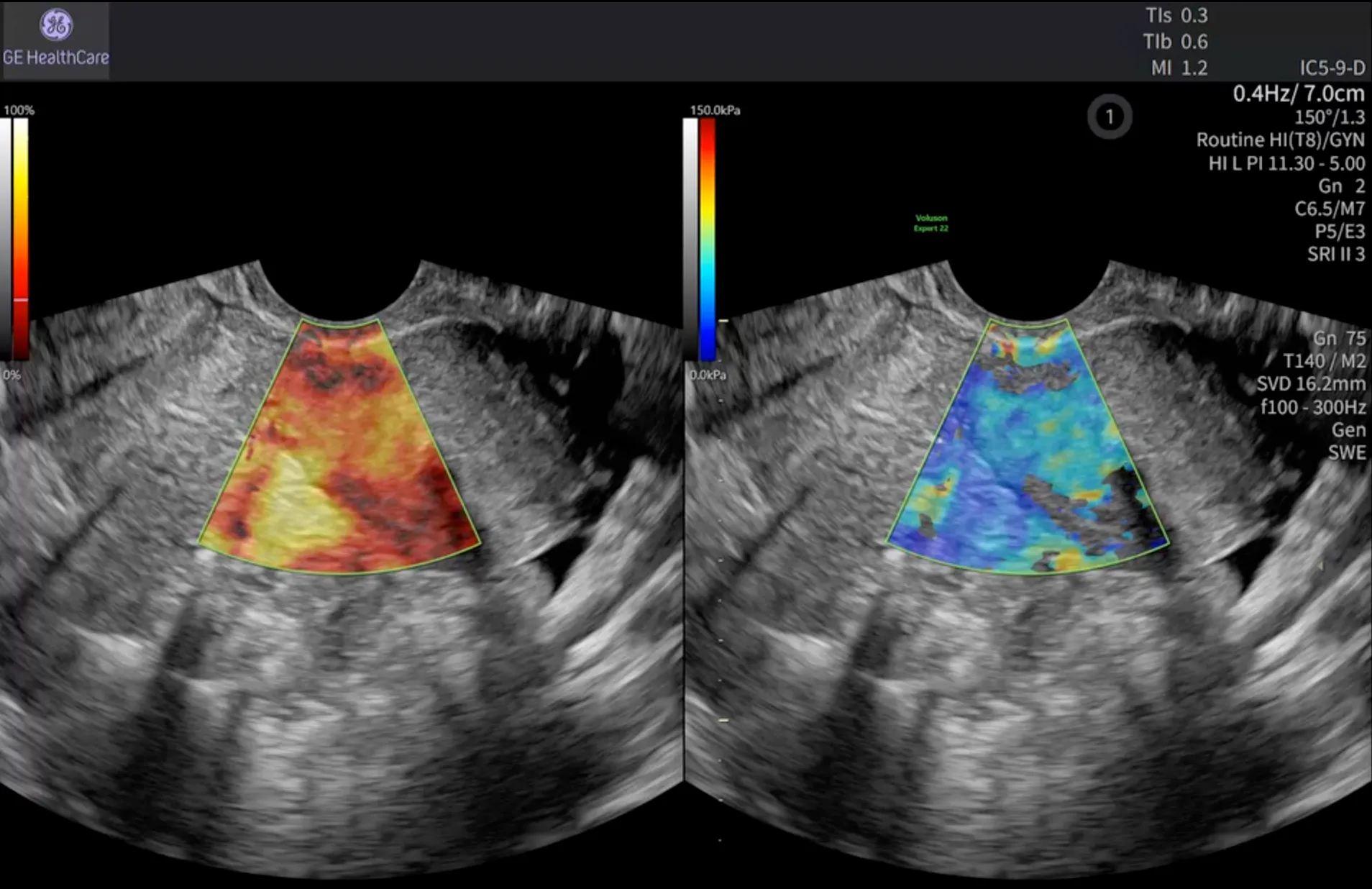
Shear Wave Elastography in Endometriosis
Revealing More Than Morphology
Not all clinically valuable information is visible on conventional ultrasound. While B-mode imaging provides an essential view of anatomy and morphology, shear wave elastography (SWE) adds quantitative information about tissue stiffness, helping clinicians gain deeper insight into suspected endometriotic lesions.¹
Endometriosis affects an estimated 10% of women of reproductive age worldwide, yet diagnosis is often delayed by seven to ten years.²˒⁴ As understanding of the disease evolves, endometriosis is increasingly recognised as a fibrotic condition. Tissue stiffness has therefore emerged as an important imaging biomarker, making SWE a valuable complement to conventional ultrasound.²
Transvaginal ultrasound remains the first-line imaging modality for evaluating suspected endometriosis.³ While conventional B-mode imaging provides critical morphological information, subtle or non-cystic disease may be more challenging to characterise. By quantifying tissue stiffness, SWE adds another dimension to image assessment, supporting a more comprehensive evaluation.¹
Advancing Endometriosis Assessment with SWE
B-mode ultrasound remains the cornerstone of endometriosis imaging, providing detailed assessment of lesion morphology and pelvic anatomy. Colour Doppler contributes vascular information, while SWE enables quantitative evaluation of tissue stiffness that may reflect underlying fibrosis.¹˒³˒⁶˒⁹
Together, these complementary technologies provide a more complete picture of disease characteristics than any single modality alone.
To achieve the greatest clinical value, SWE findings should always be interpreted in conjunction with B-mode morphology, Doppler assessment when appropriate, and the patient's clinical presentation. Although increased stiffness may suggest fibrosis, current evidence does not support universal diagnostic thresholds. As a result, stiffness measurements should be considered within the broader clinical context rather than in isolation.⁶˒⁷˒⁸
Like all advanced ultrasound technologies, SWE delivers its greatest value when combined with clinical expertise and thoughtful interpretation.
Understanding the Science Behind SWE
SWE uses focused acoustic pulses to generate shear waves within tissue and measures the speed at which those waves travel. Because shear waves move faster through stiffer tissue, clinicians can obtain quantitative information about tissue elasticity.¹
Since endometriotic lesions frequently contain a significant fibrotic component, increased tissue stiffness may provide additional information that supports lesion characterisation.²˒⁵˒⁶
Point SWE measures stiffness within a single sampling region. In contrast, 2D SWE generates a real-time, colour-coded map of tissue stiffness superimposed on the B-mode image. Because pelvic lesions are often heterogeneous, 2D SWE can help visualise regional variations within a lesion before selecting areas for quantitative measurement.¹˒⁵
Optimising Image Acquisition
Obtaining reliable SWE measurements begins with consistent image acquisition and scanning technique.
Transducer Selection
A high-frequency transvaginal transducer is recommended, providing the spatial resolution needed to assess the uterus, ovaries, and deep pelvic structures.³
Patient Preparation and Examination Workflow
No preparation beyond a standard transvaginal ultrasound examination is required. SWE should be performed following the conventional B-mode assessment so tissue stiffness can be interpreted alongside lesion morphology and pelvic anatomy.³
ROI Placement
The region of interest should be positioned within the most representative solid component of the lesion while avoiding bowel gas, large vessels, and areas affected by excessive probe pressure. Consistent and gentle transducer contact helps minimise artefacts that may influence measurements.⁵
Measurement Strategy
Multiple acquisitions should be obtained, with representative mean stiffness values used for interpretation rather than a single measurement. Because standardised reference values for gynaecologic elastography remain limited, consistency of acquisition and correlation with B-mode findings are particularly important.⁵˒⁷
Technical Limitations
As with any imaging technique, image quality remains critical. Motion artefact, bowel gas, lesion depth, and excessive transducer pressure may influence stiffness measurements and should be considered during acquisition and interpretation.⁵˒⁷
Ultimately, SWE findings should always be integrated with the complete ultrasound examination. While increased stiffness frequently reflects a greater fibrotic component, stiffness values are most meaningful when evaluated alongside conventional imaging findings and the overall clinical picture.⁶˒⁷˒⁸
Why Endometriosis Is Well Suited to Elastography
Deep infiltrating endometriosis (DIE) nodules and endometriomas are characterised by fibrosis, smooth muscle metaplasia, and inflammatory infiltration, resulting in tissue that is typically stiffer than surrounding structures.²˒⁷
This characteristic makes endometriosis particularly suited to elastographic assessment.
Clinical studies have demonstrated meaningful correlations between elastography findings and histopathology.⁶˒⁷ Increased stiffness has been associated with greater fibrotic content, with reported diagnostic sensitivities ranging from 78% to 100%.⁶ By evaluating tissue stiffness within structures such as the rectovaginal septum, uterosacral ligaments, and bowel wall, SWE may provide additional information regarding disease extent and fibrosis prior to surgery.³˒⁶˒⁹
For clinicians planning surgical intervention, understanding the location of DIE lesions, their extent, and the degree of fibrosis present is essential. By adding quantitative tissue stiffness information to conventional ultrasound findings, SWE may support more confident lesion characterisation and contribute to surgical planning.³˒⁶˒⁹
Expanding Clinical Confidence Through Comprehensive Evaluation
Despite increasing awareness, diagnostic delay remains one of the greatest challenges in endometriosis care. Improving the non-invasive characterisation of suspected lesions may help support earlier and more informed clinical decision-making.
SWE is most powerful when integrated into a comprehensive ultrasound examination. Combined with transvaginal ultrasound, Doppler assessment, and sonovaginography when indicated, SWE contributes valuable tissue information that can enhance diagnostic evaluation and treatment planning.³˒⁹
Ultimately, every ultrasound exam generates valuable clinical information. By combining quantitative tissue stiffness assessment with conventional imaging findings, SWE can help clinicians gain a more complete understanding of suspected endometriosis and support informed clinical judgement.³˒⁵˒⁶˒⁷
References:
1.Dewilde K, et al. Elastography in ultrasound assessment of the uterus. J Endometriosis Uterine Disord. 2023;2:100014. doi:10.1016/j.jeud.2023.100014
2.Ochoa Bernal MA, Fazleabas AT. The known, the unknown and the future of the pathophysiology of endometriosis. Int J Mol Sci. 2024;25(11):5815. doi:10.3390/ijms25115815
3.Guerriero S, Condous G, van den Bosch T, et al. Systematic approach to sonographic evaluation of the pelvis in women with suspected endometriosis, including terms, definitions and measurements: a consensus opinion from the International Deep Endometriosis Analysis (IDEA) group. Ultrasound Obstet Gynecol. 2016;48(3):318-332. doi:10.1002/uog.15955
4.Breton Z, Gouesbet S, Indersie E, et al. Endometriosis diagnostic delay and its correlates: results from the ComPaRe-Endometriosis Cohort. J Womens Health. 2026;35(2):172-188. doi:10.1177/15409996251380129
5.Shiina T, Nightingale KR, Palmeri ML, et al. WFUMB guidelines and recommendations for clinical use of ultrasound elastography: Part 1: Basic principles and terminology. Ultrasound Med Biol. 2015;41(5):1126-1147. doi:10.1016/j.ultrasmedbio.2015.03.009
6.Brunelli AC, Brito LGO, Moro FAS, Jales RM, Yela DA, Benetti-Pinto CL. Ultrasound elastography for the diagnosis of endometriosis and adenomyosis: a systematic review with meta-analysis. Ultrasound Med Biol. 2023;49(3):699-709. doi:10.1016/j.ultrasmedbio.2022.11.006
7.Horwood G, Flaxman T, McInnes M, McLean L, Singh SS. Ultrasound elastography in benign gynecology: a scoping review. Reprod Sci. 2024;31(8):2508-2522. doi:10.1007/s43032-024-01535-6
8.Stoelinga B, Hehenkamp WJK, Brölmann HAM, Huirne JAF. Elastography of uterine pathology: a systematic review and proposal for a standardized reporting system. Ultrasound Med Biol. 2020;46(8):1941–1955. doi:10.1016/j.ultrasmedbio.2020.04.017
9.Leonardi M, Condous G, Fedele L, et al. Ultrasound for endometriosis: moving toward a structured approach. Ultrasound Obstet Gynecol. 2020;56(3):323-331. doi:10.1002/uog.22100

