SonoAVC™follicle: Increase Exam Consistency and Improve Workflow Efficiency
SonoAVC™follicle has introduced a new level of precision to reproductive health technology that is revolutionizing how in vitro fertilization (IVF) practices care for patients. IVF success depends on many factors, and precise timing of hCG administration is one of the most critical. The days of manually counting and measuring follicles are gone, ushering in a new era of IVF care with automated tools that significantly impact clinical efficiency, patient satisfaction, and IVF success.
How 3D Technology Can Aid Clinicians
Before 3D imaging and tools like SonoAVC™follicle became available, clinicians relied on 2D imaging to assess and measure follicles during hyper-stimulation cycles. Estimating the volume of pressed together and compressed follicles takes quite a bit of skill. Also, these follicles sometimes lack clear borders, leading sonographers to wonder, Is that one mature follicle or two pressed together?
With 2D technology, it's unlikely that two different sonographers could produce the same results in the same patient on the same day. This discrepancy isn't caused by a lack of skill but by the limited information that the technology provides the sonographer.
If we consider instead how 3D technology can aid in the process of counting and measuring stimulated follicles, we understand that with appropriate tools, two sonographers are very likely to be able to produce the same results in the same patient on the same day. This higher level of consistency occurs because SonoAVC™follicle performs the calculations—not the person. This tool reduces exam time and standardizes the exam through the automated multi-planar approach, driving efficiency. Additionally, on a basic level, 3D technology means that every patient in a practice has the same paperwork and documentation in their chart, improving operational effectiveness.
How Exactly Does SonoAVC™follicle Work?
SonoAVC™follicle is, simply put, an automatic follicular assessment tool. It enhances the process of counting and measuring stimulated follicles during a 3D ultrasound.
To begin using this tool, the sonographer selects the button for SonoAVC. After they locate the area to image, they should adjust the volume angle to visualize the entire ovary. They can reduce or increase the volume angle if the field is too wide or narrow. The sonographer can also adjust the region of interest (ROI) box by touching the screen or using the machine's tracking ball.
To ensure that only the ovary is being imaged, the user should place the box's boundaries as close as possible to the ovaries while keeping the whole ovary within the ROI box. As the screen displays the follicles, the sonographer can measure them manually, semi-automatically, or fully-automatically.
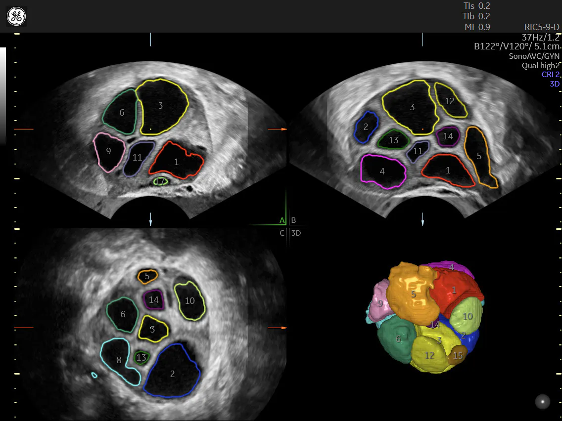
SonoAVCfollicle automatically calculates follicle volume and dimensions
Auto Caliper—tap follicle to measure
With any of the three modes, each measured follicle is listed on the right side of the screen, with the largest follicle at the top. SonoAVC™follicle can outline or completely fill each follicle with color to better visualize size and geometric shape. Clinicians can measure follicles with either an ellipse or a double caliper, depending on the user's preference. The user should deselect any non-follicle tissue picked up on the scan.
SonoAVC™follicle
SonoAVC™antral
SonoAVC™antral
As the sonographer shifts through the ovary, the follicles they have already measured remain colored, so they don't mistakenly measure them twice. However, they can remove the measurement from a single follicle and re-measure it for any reason.
At any time, the user clicks "Add to Report" to update the report. The report displays the follicles' dimensions in three ways: as a mean diameter, a volume, and a relaxed follicular diameter (which is what the diameter would be if it were not compressed by other tissue).
The most significant benefit of the fully-automated version is that it is the quickest. Some users will want to use the manual or semi-automated versions when measuring only specific follicles. Other benefits in the semi- and fully-automated versions include tools like Cut and Merge, which allow the user to separate or join follicles, or Grow, which can be used to adjust a follicle's outline if the tool appears to be measuring incompletely. Additionally, the Separation tool allows the user to separate follicles that are joining or to re-join a single follicle that appears on the screen to be separating. Increasing the separation is sometimes helpful when the machine isn't differentiating between individual follicles.
Better Patient Care with SonoAVC™follicle: Convenience, Ease-of-use, and Flexibility
SonoAVC™follicle uses volume ultrasound, eliminating the need for individual follicle measurements at the bedside, as they can be done any time after the volume is captured. Volume ultrasound captures a multidimensional model of the anatomy being imaged (in this case, the ovary), so clinicians can view it in any plane. While patients may want to see what is happening with their follicles, sometimes they appreciate that their frequent appointments can be kept brief.
This 3D reproductive health technology represents a leaps-and-bounds improvement over 2D in terms of efficiency and reproducibility when every scan counts. The timing of IVF injections no longer depends on different sonographers calculating the same follicles' measurements or even the same sonographer doing calculations on different days. SonoAVC™follicle clearly displays the shape of each follicle, automatically calculates its volume, and shows the largest follicles. This tool removes the guesswork and margin for error, increasing the likelihood of optimal hCG timing and successful pregnancies.
Interested in learning more?
Read The important role of AI and Automation in fertility care.

