The Benefits of Voluson SonoLyst AI Technology in Ultrasound Imaging
In today's rapidly evolving landscape, AI technology is seamlessly integrating into our daily lives, revolutionizing a number of sectors and significantly transforming healthcare. Particularly within ultrasound imaging and obstetric care, AI is emerging as a groundbreaking force, reshaping diagnostic imaging capabilities, ultrasound training, and patient care standards.
Professor of Fetal Medicine and Director of Research of the Oxford Maternal and Perinatal Health Institute (OMPHI), Aris Papageorghiou, underscores the pivotal role of AI technology in ultrasound exams, emphasizing its immense potential to revolutionize healthcare outcomes for women. SonoLyst, an AI algorithm, is at the forefront of this technological wave, with advanced tools that not only redefine the capabilities of healthcare professionals, but also place a heightened focus on patient care. This convergence of expertise and innovation stands poised to elevate ultrasound imaging, empower healthcare providers, and enhance the quality of care delivered to women around the world.
Image Recognition Technology from SonoLystIR
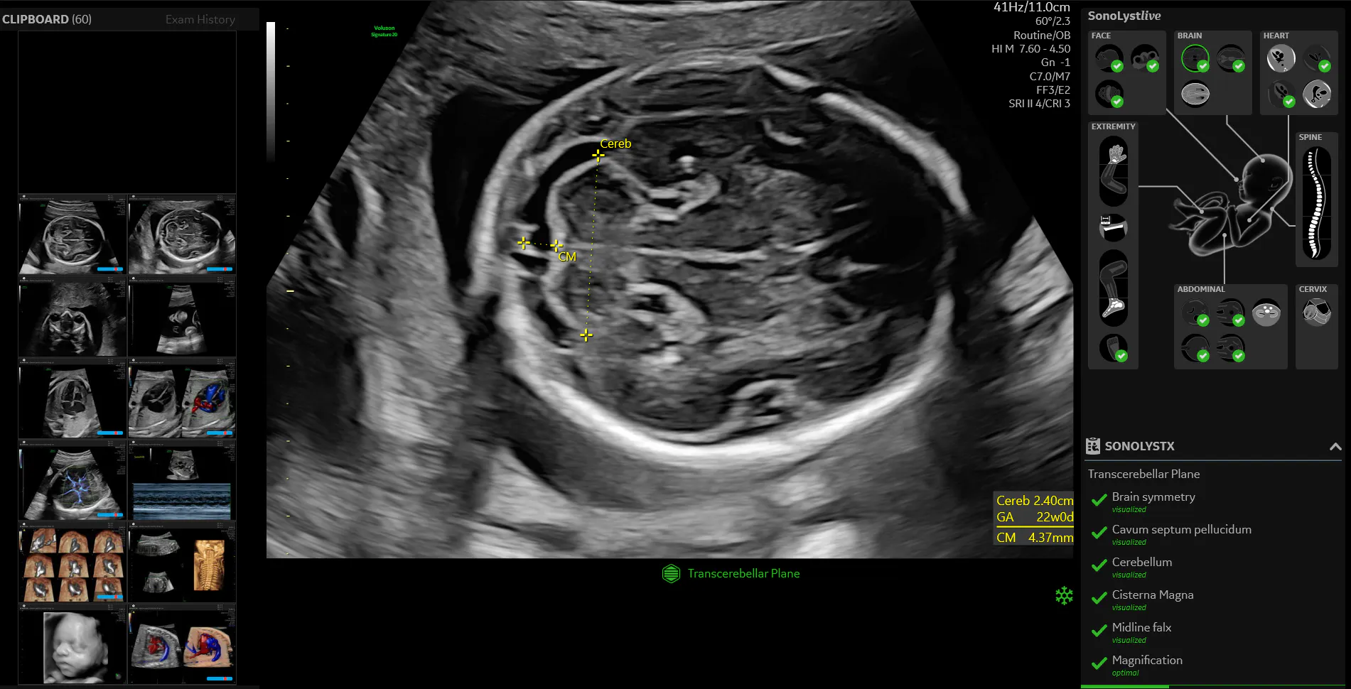
SonoLystIR (Image Recognition) revolutionizes the identification of standard fetal anatomy views in ultrasound exams with its cutting-edge image recognition technology. SonoLystIR has a focus on streamlining the recognition of ultrasound images to ensure adherence to established guidelines from the International Society of Ultrasound in Obstetrics and Gynecology (ISUOG). This tool enhances the completeness and quality of imaging exams by assisting in the identification of required views and structures as per the ISUOG guidelines and quality metrics. SonoLystIR displays a checklist and progress bar on the screen to assist operators with adherence to protocol, image quality standardization, and consistency.
Through the SonoLystIR functionality, the process of sonographic ultrasound imaging becomes remarkably streamlined: Operators assess an area or structure, select an image to freeze, and the technology swiftly takes over. SonoLystIR automatically identifies the fetal anatomy present in standard obstetrical views and performs annotating and measuring functions on the anatomical structures seen on the screen.
The tool also evaluates each image by comparing it to the accepted clinical criteria listed on the display panel, which helps to ensure that it meets the required criteria for quality. If the image is lacking in any area, the tool prompts the operator to take another image with improved optimization. This method significantly reduces exam variability between operators, ensuring a consistent and standardized approach across examinations while greatly enhancing workflow efficiency.
Moreover, SonoLyst contributes to skill building by providing comprehensive training tools, including anatomy diagrams and image examples, in order to foster continuous improvement in ultrasound proficiency and maintaining high-quality imaging standards. This combined capability of SonoLystIR and SonoLystX represents a monumental leap in ultrasound technology, promising not only precision and reliability in diagnostics but also skill refinement among healthcare professionals.
Automation and Efficiency in Ultrasound with SonoCNS
SonoCNS (Central Nervous System) is another AI-driven tool that assists in diagnostic ultrasound. It primarily specializes in CNS imaging in obstetric examinations. SonoCNS is tailored to aid in the assessment of fetal brain structures and anatomy, a very complex portion of the obstetric ultrasound exam.
SonoCNS is trained using a large number of ultrasound images and volumes, and is capable of identifying and differentiating between multiple standard views of the fetal brain. The ultrasound operator only needs to capture a 3D volume of the fetal head and SonoCNS is then activated to identify the required planes of view, discern structures such as ventricles, choroid plexus, thalamus, CSP and cerebellum. Without prompting, SonoCNS separates out the standard image planes of the brain from the volume and performs the required measurements on the choroids, cerebellum, BPD (biparietal diameter), and HC (head circumference).
The addition of this tool to the obstetric ultrasound scan routine has been found to reduce keystrokes by up to 82% and dramatically increase the reproducibility of images between operators. This represents a significant stride in streamlining the assessment and documentation of fetal anatomy by simplifying the standard routine.
This innovative tool could revolutionize workflow for healthcare practitioners and clinicians. By leveraging advanced automation and artificial intelligence programming, SonoCNS will be able to expedite the ultrasound examination process, as well as remarkably enhance the quality and reproducibility of exams, ensuring consistent and accurate results across different scans.
The Virtual Ultrasound Expert Assistant: SonoLystX
In comparison to SonoCNS and SonoLystIR, SonoLystX is a broader AI tool that acts as a knowledgeable ultrasound expert by offering real-time guidance, instructions for users on optimal transducer placement, and technique recommendations to obtain quality images.
This functionality serves a dual purpose: First, it plays a crucial role in training. It aids novices and trainees with support in mastering the complex details of ultrasound imaging by providing step-by-step assistance and feedback throughout the ultrasound exam. Secondly, SonoLystX acts as a steadfast assistant for experienced professionals, ensuring ongoing adherence to high quality imaging standards, which bolsters quality assurance efforts within the healthcare setting.
Think of SonoLystX as a virtual mentor offering guidance and support to ultrasound operators that not only facilitates skill enhancement, but also ensures the preservation and management of exemplary imaging standards across various healthcare situations.
The Future of AI in Ultrasound
The SonoLyst technology is likened to that of having a virtual on-board assistant. It utilizes the power of AI to identify fetal anatomy from standard obstetrical ultrasound views while enhancing efficiency of the scan routine with the addition of annotations, measurements, and quality metrics. And you can save up to 40% on routine second trimester exams with SonoLyst. AI tools have the power to transform second trimester obstetrical ultrasound exams, which are notoriously complex and detailed examinations, where accuracy, precision, and reproducibility are critical components of a properly completed assessment.
AI technology has tremendous potential for revolutionizing healthcare in several ways. Algorithms can enhance diagnostic accuracy by continuously evolving to become more adept at analyzing ultrasound images. AI can assist in identifying subtle anomalies or patterns that might be missed by human eyes, thereby improving diagnostic accuracy and early disease detection.
AI tools, like SonoLyst, can also help optimize workflow by streamlining ultrasound examinations and automating certain processes, reducing the time needed for scans. This efficiency benefits both healthcare providers and patients by expediting diagnosis and enabling more patients to be seen in less time. There is also the possibility that personalized healthcare is a future option with AI-driven ultrasound by providing imaging and examinations that contain more precise and tailored diagnostic information. This could lead to more individualized treatment plans and better patient outcomes.
Additionally, AI tools in ultrasound imaging can serve as valuable educational aids for training new healthcare professionals and further educating the experienced ones. These tools offer guidance and feedback, helping individuals learn and improve their skills in understanding and interpreting ultrasound images.
The integration of AI is anticipated to evolve, offering real-time analysis, predictive capabilities, and nuanced insights into complex imaging data, propelling ultrasound technology into an era of unparalleled diagnostic capability and transformative patient care. Papageorghiou envisions AI as a catalyst that not only accelerates the path to diagnoses, but also increases the precision of imaging, elevating healthcare to new levels. As technology advances and collaborations between AI developers and healthcare providers continue, the future of AI technology in ultrasound holds immense promise, with applications that will continue to improve and perhaps even revolutionize the field of medical imaging.
Interested in viewing lectures about the power of AI? Click below:
Professor Aris Papageorghiou – AI power for obstetric imaging.
Megan Meade – Voluson AI in fetal anatomy exam.

