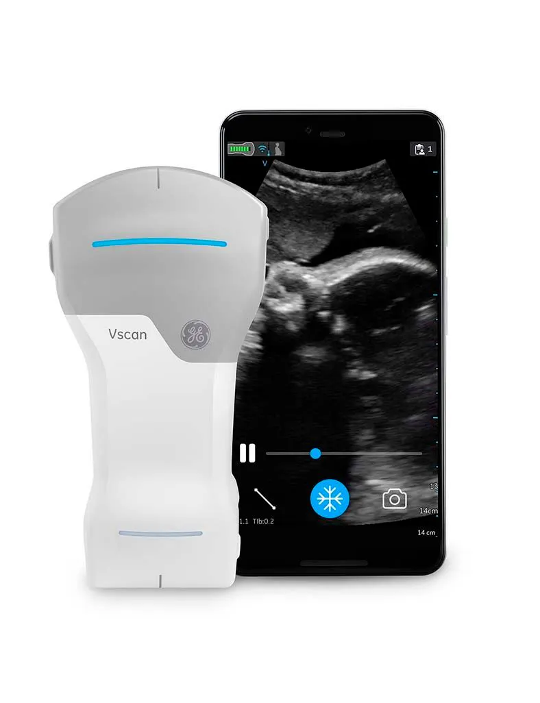
The Role of Handheld Ultrasound in Quick-Check Obstetrics
Handheld ultrasound is revolutionizing the practice of imaging in obstetrics. There are many uses for this new technology in obstetrics: from determining viability to dating the pregnancy to identifying singleton or multiple pregnancies.
Handheld ultrasonography can also ascertain the location of the placenta, rule out placenta previa, determine fetal presentation and fetal position prior to instrumental delivery, identify any abnormalities of amniotic fluid volume, and diagnose whether the fetus has a structural abnormality. In early pregnancy, physicians may use it to determine pregnancy viability. The ease of access to handheld devices, their lower cost, and the fact that they are easy to use makes them attractive for community practices in low- and middle-income countries. As the technology has improved over the years, the quality of the ultrasound images obtained using handheld devices is approaching or equal to the quality of traditional ultrasound systems.
One systematic review of 16 studies found that scans produced with these devices are comparable to the high-end ultrasound systems, that they are a valuable supplement to a physical examination performed by an obstetrician, and that they have the potential to become a complementary and accessible diagnostic tool for obstetric patients.
Identifying Breech Presentation with Handheld Ultrasound
While it's true that fetal presentation assessment is important at term, it's also important earlier in pregnancy, particularly for a woman who presents with preterm labor or premature rupture of membranes, according to Dr. Asma Khalil, professor of obstetrics and maternal fetal medicine at St. George's Hospital, University of London.
The incidence of breech presentation varies according to gestational age, says Khalil, tending to be more common earlier in the third trimester. "The problem with breech presentation is that it's associated with excess perinatal mortality and morbidity and also has medicolegal implications," says Khalil.
When a diagnosis of antenatal breech presentation is made near term, a clinician should discuss three options in detail with the pregnant person: external cephalic version or ECV, planned cesarean section, or planned vaginal breech birth.
Universal Late Pregnancy Ultrasound Eliminates Undiagnosed Breech Presentation Labor
Because a third trimester ultrasound is not standard in the UK (though some practices do offer it), 30% to nearly 50% of babies presenting breech are only detected when the mother is already in labor, according to Khalil. At that point "there are only two options," she says: emergency cesarean section or unexpected vaginal breech delivery. "Both options carry maternal and infant risks," says Khalil.
What about offering universal late pregnancy ultrasound? Universal late pregnancy ultrasound eliminates undiagnosed breech presentation labor and therefore reduces the risk of perinatal mortality and morbidity. "It's estimated that it saves at least seven babies' lives a year across the United Kingdom," says Khalil. "And, of course, that means we can offer an informed choice of model delivery to the pregnant woman."
Cost-Effectiveness of Handheld Ultrasound Scans
According to the UK figures, the universal late third trimester ultrasound scan would be cost-effective if the cost is less than £20, says Khalil. However, the national tariff ultrasound scan performed by a sonographer in the United Kingdom is just under £50. Therefore, doing a traditional third trimester ultrasound scan is not cost-effective.
"However, if we are able to use a low-cost handheld device rather than the high-end ultrasound systems and have the midwife or physician perform the scan during a routine third trimester visit, that means that we could potentially meet the threshold for cost-efficiency," says Khalil.
Performing a routine third trimester presentation scan with a handheld device has the potential to improve clinical outcomes. From the service point of view, access to ultrasound is improved: waiting times are reduced, and the pregnant person does not have to travel to the hospital for the scan. "This can be important, particularly in healthcare systems or countries where there is a shortage of sonographers," says Khalil.
Use in Obstetric Emergencies
Formal studies that assess how much time is saved by using handheld ultrasound in an obstetric emergency have yet to be conducted, says Khalil, but her team did a very simple experiment: they timed how long it took to turn on a standard ultrasound machine versus the handheld one. The handheld one took only 35 seconds, compared with 2 minutes and 20 seconds for the standard device.
Though more research is needed, handheld devices have the potential to save time in obstetric emergencies.
Looking to the Future
Khalil is optimistic about the role of handheld ultrasound in obstetrics. While healthcare professionals will need to complete training and ensure competency in this new modality, the potential for handheld devices to provide easier access to ultrasound is significant. In particular, their use in low- to middle-income countries and in the community setting could be very useful, Khalil says. Pregnant people who have experienced this type of scan have indicated a generally positive perception. They cited the ease of access, especially not having to go to the hospital for the scan, as well as the reassurance of knowing their baby's presentation.
In the future, as healthcare becomes more digital and technology more advanced, the use of devices like handheld ultrasound will likely become more commonplace.
View Professor Asma Khalil’s video here: Point of care ultrasound in obstetrics.

