Transperineal Ultrasound: From the Pelvic Floor to the Labor Ward - Meet the Expert, Dr. Aly Youssef
Join Dr. Aly Youssef for a presentation on the use of transperineal ultrasound for both the pelvic floor and labor and delivery. Dr. Youssef is the author of "The maternal pelvic floor and labor outcome" and ISUOG Practice Guidelines intrapartum ultrasound, 2018. He is also a member of the International Society of Ultrasound in Obstetrics and Gynecology (ISUOG), with over 70 published articles on the topic of ultrasound assessment of the female pelvic floor.
The transperineal approach is the use of ultrasound from the perineal or labial approach to assess the muscles of the pelvic floor and the process of labor. Transperineal ultrasound has allowed for a better understanding of the direct link between female pelvic floor dysfunction/damage and vaginal deliveries.
One of the main requirements of a successful vaginal delivery is the maternal ability to effectively push, which is the relaxation of the levator ani muscle (the primary pelvic floor muscle). Transperineal ultrasound has the ability to assess both the dynamic and static conditions of the levator ani muscle. Longer durations of labor and higher risk of cesarean deliveries, as well as increased stress on the mother and fetus are associated with absent or incomplete levator ani muscle relaxation and smaller overall levator ani hiatal size. Transperineal ultrasound has been found to be an effective and reproducible method to image and assess the levator ani muscle to determine if damage or dysfunction is present and to help determine labor outcome.
Applications in Pelvic Floor Assessment
Pelvic floor transperineal ultrasound can be used to evaluate the integrity and function of the pelvic floor three to six months after delivery. Ultrasound can identify tears and levator ani muscle injuries, which affect pelvic health.
Two types of lesions can affect the levator ani muscle:
- Macrotrauma (avulsion) injury. The detachment of the levator ani muscle from its insertion in the pubic symphysis.
- Microtrauma (overdistension) injury. Occurs where the levator ani muscle has lost its tone and is overdistended but not damaged macroscopically.
Consequences of macrotrauma and microtrauma to the levator ani muscle include reduced activity of the pelvic floor and increased risk of prolapse of the pelvic organs.
SonoPelvicFloor - Voluson Expert 20
SonoPelvicFloor- Voluson Expert 20, RAB6
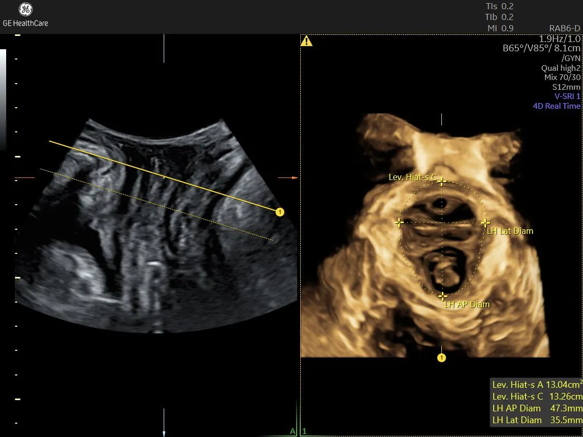
Clinicians can assess the levator ani muscle with 3D ultrasound, which allows for reconstruction of the plane of minimal hiatal dimensions, or the angle that the levator ani muscle runs from the pubic symphysis to the anorectal angle. The image reconstructed by this plane on 3D ultrasound includes visualization of the pubis, the urethra, the vagina, the rectum, and the area included inside the levator hiatus.
More detailed information on pelvic floor muscle contraction can be gained by the use of linear reconstruction with contrast enhancement. This technique is done at rest, during contraction, and during pushing (Valsalva maneuver). If normal, the levator hiatus dimensions should reduce during contraction and increase during Valsalva.
SonoPelvicFloor - Voluson Expert, RM7c
SonoPelvicFloor - RM7c probe
The multislice technique can also be applied to identify areas of the levator ani muscle that are intact, showing signs of avulsion, or microtrauma (seen as a ballooning area > 25 sq cm on Valsalva).
Ultrasound assessment done three to six months after delivery can help diagnose any potentially missed perineal tears post vaginal delivery. Translabial ultrasound is used in the axial plane, and both the internal anal sphincter and external anal sphincter can be identified. Signs of damage to the internal, external, or both areas of the anal sphincter are seen by looking for a loss of the "star sign," visual defects, and the half moon sign.
Implications for the Labor & Delivery Ward
Levator ani assessment can be done by looking at the antero-posterior diameter (APD) of the levator hiatus from the transperineal approach. Ultrasound can demonstrate the pubic symphysis, urethra and bladder, vagina, rectum, and puborectalis muscle. In 2D ultrasound, the sagittal view of all of these structures represents the antero-posterior diameter (APD) of the levator hiatus. A cine video taken in this plane can demonstrate the contractility of the levator ani muscle. Good contractility will show the rectum being pulled anteriorly toward the pubic symphysis.
Transperineal ultrasound can also document the movement of the pelvic floor muscles with a Valsalva maneuver, when the pelvic floor should relax. During relaxation, the bladder should move inferiorly (toward the transducer) and the levator ani muscle should move posteriorly (toward the right of the screen). The ability to relax the maternal pelvic floor is crucial for vaginal delivery, when the muscles of the pelvic floor must be able to relax enough to allow the fetal head to pass through. This is seen on ultrasound as an increase in the levator hiatus APD.
Some women are unable to relax their pelvic floor during a Valsalva maneuver, which reduces the APD of the levator hiatus, which is known as levator ani co-activation. There is a consistent association between smaller levator hiatal dimensions, levator ani co-activation, and increased duration of the second stage of labor.
Intrapartum Ultrasound Exams
Clinicians can use two methods to conduct intrapartum ultrasound exams:
- Transabdominal approach. A systematic examination of the fetus in labor should always begin with a transabdominal ultrasound exam to determine the position of the fetal head.
- Transperineal approach. Used to assess fetal head descent and rotation.
These approaches allow providers to assess two parameters related to the progression of labor:
- Angle of progression (AoP). A measurement taken in the sagittal position from an angle between two lines. The first line is through the long axis of the pubic symphysis bone and the other line extends from the most inferior point of the pubic symphysis to pass tangentially across the most inferior aspect of the fetal skull. The more inferior the fetal head in the birth canal, the wider the angle of progression will be and more imminent the vaginal delivery.
- Head perineum distance (HPD). A measurement taken in the axial (or transverse view) by rotating the ultrasound transducer 90 degrees anticlockwise from sagittal, which provides an axial view of the maternal birth canal and fetal skull. The clinician measures the distance between the fetal skull and transducer, and the smaller the distance, the more engaged the fetal head is in the birth canal.
One of the most important uses of the HPD measurement is in cases where an instrumental delivery is being considered. If the fetus is in the occiput-anterior position with an HPD ≤ 35mm, the success rate of instrumental delivery is approximately 98%. And if the AoP is ≥ 146 degrees at rest or ≥ 154 degrees with pushing, these indicate very good rates of success for an instrumental delivery. The ISUOG Practice Guidelines for intrapartum ultrasound summarize the complete information on how to perform intrapartum ultrasound exams.
Long-Term Benefits of Transperineal Ultrasound for Women
Transperineal ultrasound has demonstrated an ability to bring significant amounts of detail to light about the female pelvic floor. Dr. Youssef notes that ultrasound does no harm to the patient, so it is always worth attempting for the extra information it can provide. Transperineal ultrasound imaging can also be integrated into regular patient care to help women understand how to better contract or relax their pelvic floor muscles and correct any errors in function.
In general, transperineal ultrasound has been found to be an objective and reproducible tool for pelvic floor function, anatomy, and fetal head descent during labor. We know that pregnancy and delivery are the most common causes of pelvic floor injury. Follow-up assessments utilizing transperineal ultrasound can improve recovery and prevent future risk of long-term damage to the levator ani muscle and pelvis organ prolapse. In this way, transperineal ultrasound has the ability to improve the overall female experience, both during and after labor and delivery.

