

As patients evaluate options for the treatment of uterine fibroids, they will seek advice from their physicians, who must utilize imaging to understand each unique situation, rule out contraindications, determine the types of fibroids present and settle on the best route for intervention and follow-up care.
Ultrasound fills many of these roles, and its functionality is only expanding. Follow along to learn more about ultrasound for uterine fibroids, one of the most common female pelvic disorders.
Diagnosing, Classifying and Treating Uterine Fibroids
Routine Pelvic Exams: Most fibroids are discovered during bimanual examination or incidentally during a pelvic ultrasound. ObGyn.net recommends performing a transvaginal ultrasound whenever an abnormality is felt during an examination or when a patient has abnormal bleeding or cramping symptoms. Ultrasound can quickly help distinguish between fibroids, endometriosis, adenomyosis, ovarian cysts or other pathology — each of which lead to different treatments.
The FIGO System: The most commonly used fibroid classification program is based on the FIGO abnormal uterine bleeding system. The fibroids are categorized as submucosal, intramural, serosal or "other" based on the appearance of the fibroid ultrasound results. The fibroid positions are often used as essential guidelines to determine eligibility for ablation procedures. A diagnosis employing this system is performed by 2D and 3D ultrasound and can also include sonohysterography to clarify the submucosal location of fibroids.
3D Sonohysterography: Direct uterine cavity evaluation is often necessary to assess for intracavitary or submucosal fibroids. 3D sonohysterography uses a small amount of saline solution inserted into the uterus to clearly image the endometrium on an ultrasound. It can be easily incorporated into a routine transvaginal ultrasound exam when assessing the cause of abnormal uterine bleeding. This procedure has a diagnostic accuracy comparable to hysteroscopy in the detection of intracavitary uterine abnormalities, according to The Egyptian Journal of Radiology and Nuclear Medicine.
Radiofrequency Ablation (RFA): This minimally invasive, uterine-sparing treatment of uterine fibroids is guided by ultrasound. The accurate placement of the needle electrodes is crucial for successful treatment. Ablation guided by ultrasound can include laparoscopic, transcervical-intrauterine or transvaginal procedures.
Ultrasound Guided Hysteroscopic Myomectomy (UGHM): Fibroids located within or abutting the endometrial cavity can cause abnormally heavy bleeding. The European Journal of Obstetrics & Gynecology and Reproductive Biology reported on the safe and effective removal of submucosal or intracavitary fibroids via an ultrasound-guided resectoscope. Ultrasound is essential in monitoring the surgery in three dimensions to ensure efficient cauterization and preventing uterine perforation.
Uterine Artery Embolization (UAE): Symptomatic fibroids can be treated by vascular embolization, allowing them to shrink and reducing or eliminating related symptoms. Prior to the procedure, ultrasound helps to identify contraindications, such as pelvic malignancy or pedunculated fibroids. RadioGraphics reports that ultrasound is very effective in detecting most of the commonly recognized complications associated with embolization and monitoring the regression of fibroids.
Modern Ultrasound for Uterine Fibroids
Considering the vast improvements in ultrasound technology, it should be no surprise that a uterine fibroids ultrasound can be effective as a part of treatment. Ultrasound, in one or more of its varied forms, can be useful all the way from the initial assessment of a bulky uterus to diagnosis, classification, treatment and post-procedure follow-up.
Many patients also prefer an ultrasound, especially a transvaginal ultrasound, over other imaging modalities, making it easier than ever to use ultrasound in nearly all fibroid-related medical endeavors.
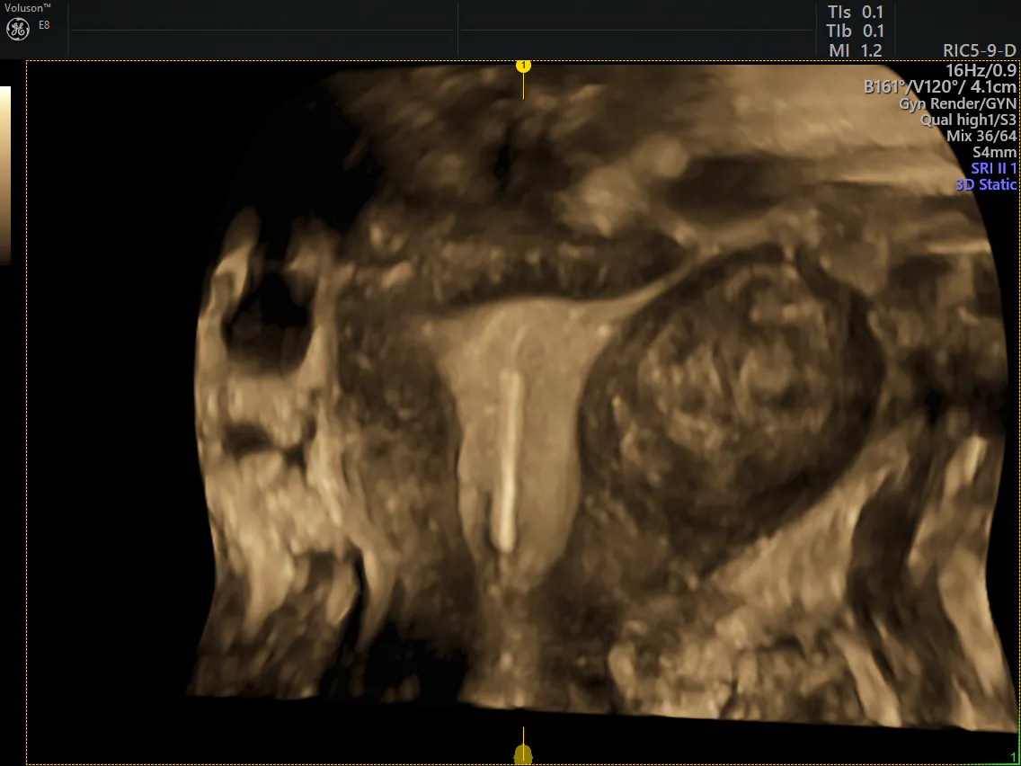
3D uterine fibroid ultrasound pictures

