

Ultrasound is one of the most useful first-line assessment tools for the female pelvis — in particular, for assessing pathology of the endometrium. Transvaginal ultrasound can easily detect a problem by measuring endometrial thickness, but a thickened endometrium can be a nonspecific finding that typically warrants further examination to evaluate suspected pathology.
Color Doppler can increase the specificity of a diagnosis by using vascular assessment to differentiate between endometrial cancer, hyperplasia, polyps and submucosal fibroids.
What Color Doppler Can Reveal About Endometrial Pathology
If a thickened endometrium is discovered, a second-line exam is typically required to further evaluate the suspected pathology.
When assessing the endometrium, transvaginal ultrasound with color Doppler can be used to provide additional information, which you cannot see using 2D ultrasound alone. Ultrasound is accessible, cost-effective and often preferred by patients over other more invasive testing.
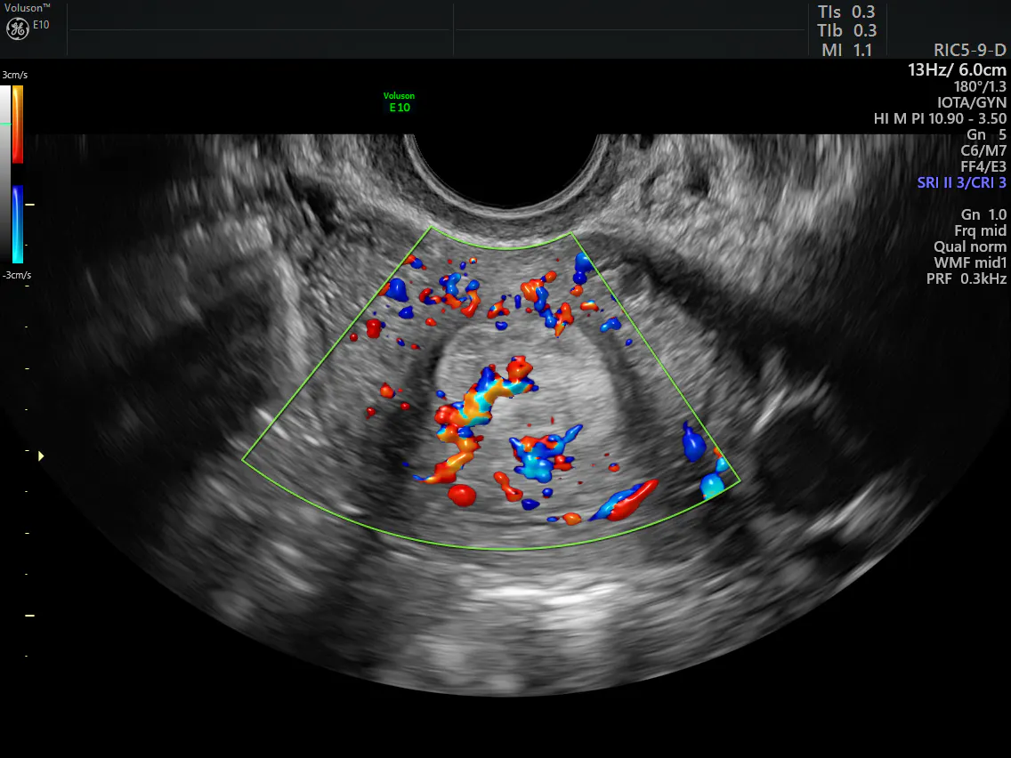
Vascular patterns can provide additional insight into the endometrium. For example, when differentiating between a polyp and submucosal fibroid — both focal endometrial lesions — the polyp will have a distinct feeding vessel, sometimes referred to as a single vessel pattern or pedicle artery sign. This vessel runs centrally through the endometrium into its base.
A submucosal fibroid has color flow in a distinct circular or semicircular pattern around its rim, sometimes accompanied by a few small, scattered vessels within. If color mapping fails to reveal clear vascular patterns within the endometrium, power Doppler may improve detection due to its increased sensitivity to low-velocity blood flow.
These distinctive Doppler findings have been well documented. In a study of 49 patients published in Ultrasound in Obstetrics and Gynecology, researchers found that 81.3% of endometrial polyps examined had a distinctive single vessel pattern visible on transvaginal sonography; 70.6% of submucosal fibroids examined had a rim-like vessel pattern.
Endometrial Hyperplasic and Malignant Lesions: Ultrasound Markers
In the higher-risk group of postmenopausal women with bleeding and a thickened endometrium, differentiating between benign endometrial hyperplasia and malignant lesions is especially critical. In this scenario, color Doppler also reveals clear distinctions in vascular patterns.
One study found that the majority of endometrial cancers presented with a multiple vessel pattern on power Doppler, while the majority of hyperplasias produced a scattered vessel pattern. Color Doppler also helps distinguish between types of malignancies. In an Ultrasound in Obstetrics and Gynecology study comparing the ultrasound characteristics of primary endometrial cancer with coexistent ovarian lesions and primary endometrial cancer with ovarian metastasis, researchers found that a multiple-vessel pattern occurred more often in endometrial cancers with ovarian metastasis than in cases without. This preoperative identification of malignancy can inform and streamline patient management.
Ultrasound's Reliability in Diagnosing Endometrial Pathology
The International Endometrial Tumor Analysis (IETA) group uses color mapping in describing the sonographic features of the endometrium — and for good reason. The Journal of Ultrasound in Medicine reports that when using color Doppler with 3D ultrasound uterine volumes in the assessment of endometrial vascular architecture, there was "very good" intraobserver and interobserver agreement, regardless of the observer's level of experience.
A study published by the European Journal of Gynaecological Oncology reports that color Doppler ultrasound results confirm diagnoses made using hysteroscopy, biopsy and curettage in more than 92% of cases when examining endometrial lesions. With its high rate of detection, transvaginal ultrasound with color Doppler is becoming a valuable tool for clinicians differentiating between endometrial pathologies.

