

The focus of gynecological ultrasound assessment has long been on transabdominal and transvaginal methods. However, there is one further ultrasound technique that also provides the ability to offer a pelvic assessment: the transperineal ultrasound.
The benefits of a superficial transperineal examination are that it does not require a full bladder or vaginal entrance. This is excellent news for patients who have difficulty with bladder filling, adolescents and even patients with a larger body habitus. Abnormalities of the vaginal canal, cervix, urethra, rectum and pelvic floor muscles can often be overlooked or shadowed out when assessed with transabdominal or transvaginal ultrasound. However, this anatomy may also be visualized via the transperineal approach, including tiny lesions in these areas that often contribute significantly to distressing symptoms.
Including a perineal assessment in the toolkit for pelvic ultrasounds can help clinicians screen a wider variety of patients.
Examination Techniques for the Superficial Transperineal Ultrasound
A transperineal ultrasound is performed with the patient in a supine lithotomy position, with the transducer sheathed in a protective covering and adequate gel used on the perineal area between the labia minora and labia majora. Because of this positioning, this technique is also referred to as a translabial ultrasound.
The transducer should be centered with the vaginal orifice located posteriorly and the urethral orifice anteriorly. Depending on the organ requiring assessment and the depth of ultrasound visualization desired, either a lower frequency curvilinear or higher frequency linear transducer can be used. Imaging planes include sagittal, oblique and coronal, with the sagittal plane being the most useful for assessing the greatest number of organs.
Imaging is typically performed with the patient in a state of rest. For some assessments, such as of the pelvic floor, imaging can also be performed during the Valsalva and pelvic floor contraction maneuvers. The use of 3D volumetric transducers can also provide an axial plane view, which is useful in the evaluation of certain pelvic floor muscles, according to research from the International Journal of Women's Health.
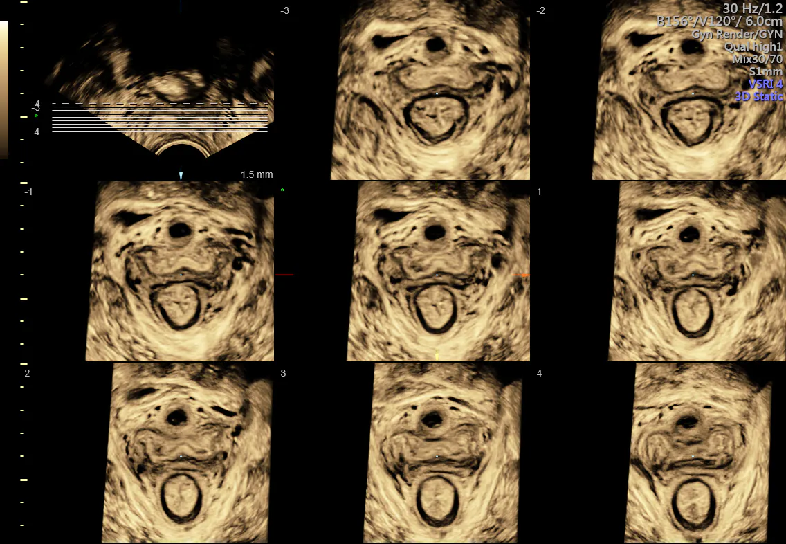
3D of normal pelvic floor in TUI display (tomographic ultrasound imaging)
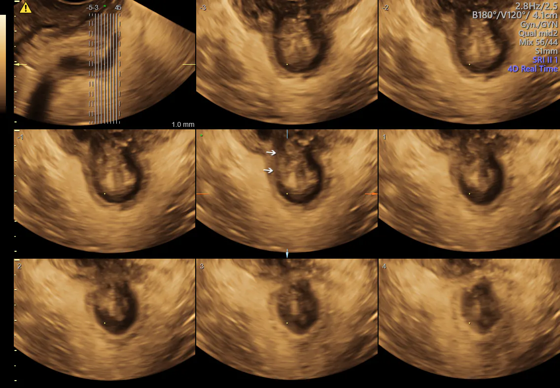
3D of pelvic floor demonstrating a tear in the external anal sphincter
Managing Vaginal and Other Lower Pelvic Pathology With Ultrasound
Transperineal ultrasound offers a significant advantage in the face of certain vaginal pathologies, such as blockage, atresia, agenesis or cysts that are present with an intact hymen. It can provide a high-resolution assessment of the entire vaginal canal, allowing for the identification of abnormal masses, lesions or transverse/longitudinal septa.
A pictorial review published in the British Journal of Radiology reports that because ultrasound exams occur in real time, the dynamic assessment allowed by the transperineal approach can effectively identify very small or subtle vaginal abnormalities. Vaginal fistulas and pelvic floor dysfunction can be clearly identified with transperineal imaging, as well as pathology posterior to the hymen, which would otherwise be obscured in the transabdominal approach and not visible transvaginally.
Transperineal ultrasound is also valuable for its ability to assess structural and dynamic pelvic floor dysfunction, according to a study published in the Middle East Fertility Society Journal. Most patients still have a limited understanding of the effects of pelvic floor dysfunction, but it has been found to be the cause of fecal and stress incontinence, pelvic organ prolapse, chronic pelvic pain and sexual dysfunction.
Most cases of pelvic floor disorder only require a correct assessment to lead to effective treatments, which may include physiotherapy or devices such as pessaries based on an individual patient's needs. OB/GYNs may want to consider a referral to a urogynecologist for treatment of pelvic floor dysfunction.
Incorporating Transperineal Imaging Into Pelvic Ultrasound Routines
The vagina, urethra and pelvic floor muscles can be easily overlooked by most imaging modalities, including transabdominal and transvaginal ultrasound. Any suspicion of vaginal pathology, or symptoms of pelvic pain without a clear cause identified through other assessments, should be considered for transperineal ultrasound evaluation.
Younger patients, adolescents or those who decline a transvaginal exam are also excellent candidates for a transperineal assessment. This scan adds no cost or inconvenience to patients already having an ultrasound exam, and it can contribute significantly useful information that can lead to effective treatment and patient management.

