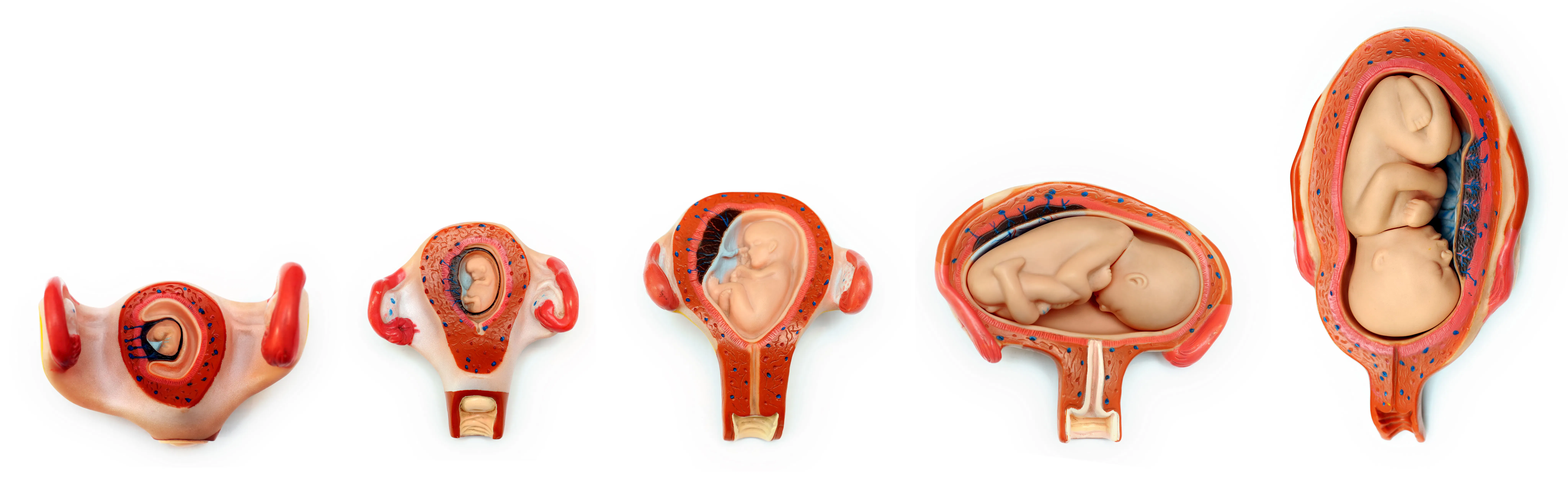
Using Ultrasound to Assess the Prenatal Brain Development Stages
Fetal brain development begins very early in the first trimester of pregnancy. In every one of the prenatal brain development stages, multiple neural structures are forming and growing. Routine obstetrical ultrasound exams are used to examine the fetal brain and surrounding anatomy to help determine if there are any early signs of abnormal neural development or function.
Fetal Brain Development Chart
As early as the third week of gestation, a neural plate forms out of a group of rapidly multiplying cells. The neural plate folds in on itself to become a tube. This neural tube then develops into the forebrain, midbrain, hindbrain and spinal cord. By the fifth week of gestation, neurons are already forming and multiplying.
During the second trimester, the majority of activity and development takes place in the fetal brain. Between the fourth and sixth months of gestation, as many as 250,000 neurons are being created every minute. These neurons start to take on specific roles and migrate to different regions of the developing brain to begin the processes involved with storing memories, interpreting sounds and connecting with other neurons.
Throughout this trimester, the development of physical reflexes occurs. The fetal brain and nerves are at the stage where the vital functions of sleeping, swallowing and breathing can be controlled as well as the physical reflexes of movement. These movements can be identified on an ultrasound exam and can be noticed by the mother as fetal kicks.
By the seventh month of gestation, the fetal cerebral cortex is growing more rapidly than any other part of the brain. The cerebral cortex contains as much as 70% of the fetal neurons and is the part of the brain where language and abstract thought function happens. The process of myelination also occurs at this stage — a dense fatty substance forms along the connections between neurons that allows signals to pass between them with greater speed.
By the eighth month of gestation, the visual cortex, auditory cortex and area of the brain associated with speech are beginning to function, which is the very beginning of cognitive development.
Ultrasound Assessment of Fetal Brain Development
In the first trimester, a dating ultrasound is typically performed at 7-12 weeks of gestational age, when the very small fetal pole can be measured, and the primitive fetal heart can be seen beating. At this stage, the fetus can already demonstrate some movement, which reveals that the brain and electrical impulses that govern movement are beginning to function.
During the 20-week detailed obstetric ultrasound in the second trimester, specific parts of the fetal body are assessed with particular care. The overall shape and size of the skull are assessed as well as certain brain components.
Skull measurements
To determine if the size of the skull is within normal limits, the biparietal diameter (BPD) and head circumference (HC) measurements are taken. The BPD is obtained from an axial plane through the fetal head at the level of the thalami and cavum septum pellucidi (CSP).
The HC is taken from an axial plane of the fetal head at the same level as the BPD. The landmarks that must be visible in this image are the CSP, thalami, falx and both choroid plexus. This measurement is made all the way around the outer edge of the bone of the skull and is part of a formula that is used to determine if the fetal skull and brain are at an appropriate size for gestational age.
If the BPD and HC measurements are smaller than typical, it can indicate that brain development is not progressing optimally or that there is an existing abnormality present in the fetus causing the size discrepancy. These two measurements are also used along with measurements of the fetal abdominal circumference and femur length to generate a complete picture of fetal size, weight and overall development. These are determined with a standard set of charts that provide values by gestational age and percentile.
Brain assessments
The specific brain components assessed during this routine ultrasound exam include the cerebellum, cisterna magna, CSP, thalami and lateral ventricles.
When assessing the cerebellum, the normal width in millimeters should correspond to the number of weeks of gestational age of the fetus (i.e., a 20-week fetus has a normal cerebellum width of approx 20 mm). If the cerebellum is abnormally small or large, it can be an indication of a variety of abnormalities, including Dandy-Walker syndrome and Arnold-Chiari malformations.
The cisterna magna is a cystic space between the cerebellum and occipital bone that also requires assessment and measurement. A normal cisterna magna has a diameter of 2-10 mm. If the cisterna magna is measuring larger than 10 mm, it can be a sign of Dandy-Walker syndrome. And if the cisterna magna is smaller than 2 mm or not visible, it can be due to some form of spina bifida.
The CSP is a structure that also needs to be confidently identified in the middle of the normal fetal brain. It can be absent in conditions such as holoprosencephaly or central nervous system malformations.
Lastly, the lateral ventricles containing the choroid plexus, found bilaterally in both sides of the fetal brain, are also assessed. The width of the ventricle atrium is normal when it is 10 mm or less. When one or both ventricles measure greater than 10 mm, it can be a sign of ventriculomegaly. This is a relatively common prenatal finding and can have many causes, such as intrauterine infection, chromosomal abnormalities or central nervous system malformation.
The choroid plexus structure of the lateral ventricle can also be affected by the development of a choroid plexus cyst. The presence of this cyst along with other fetal structural abnormalities can indicate the presence of trisomy 18 or trisomy 21.
If the cerebellum, cisterna magna, CSP and lateral ventricles can all be identified with ultrasound as present and within normal size limits, it offers reassuring evidence that the fetal brain as a whole is structurally normal. However, if any of them is found to be abnormal, further investigation needs to be done with ultrasound and other tests to determine the cause of the abnormality.
With the advancement of ultrasound technology, neurosonographic studies have found that the use of 3D ultrasound in utero is able to provide an even more detailed multiplanar assessment of the fetal brain in the second trimester. This allows better visualization of landmarks and abnormalities, ensuring for more accurate diagnosis.
Prevention, Treatment and Improvements to Fetal Brain Imaging
A healthy daily lifestyle is beneficial for the developing fetus throughout the entire prenatal brain development stages. Pregnant patients should be encouraged to engage in regular, moderate physical activity, take recommended supplements, eat a healthy and well-balanced diet and get enough rest. Research published in Science Direct shows that maternal activities and choices that lead to better nutrition, less stress and reduced rates of infection have been shown to be beneficial to the neurodevelopmental outcome of a fetus.
However, once an abnormality is identified, treatments need to be considered. The treatments available will vary depending on the severity and type of condition present. Many focus on managing specific symptoms. Indications of seizures can be treated with anticonvulsant medications. Other conditions can be treated with surgery. In hydrocephalus, the insertion of shunts is used to drain excess cerebrospinal fluid, and surgical modification of malformed skulls can allow space for the brain to grow normally.
Unfortunately, abnormalities in brain development often have severe repercussions for the fetus. Some forms of intellectual deficiency or disability can occur if the condition is mild, and if the condition is severe, it can lead to fetal death.
Earlier identification of fetal brain abnormalities can allow for more time to determine a course of treatment and more options for the parents. Most brain abnormalities are present in the first trimester of pregnancy, but the majority are not identified until the second trimester due to the small size of the fetal cranium and an assessment limited to the cranial bones, midline falx and lateral ventricles.
With the use of multiplanar neurosonography and 3D ultrasound, additional brain details have been found to be visible at 11-13 weeks, allowing for abnormalities such as ventriculomegaly, open spina bifida, Dandy-Walker syndrome and corpus callosum agenesis to be identified in the first trimester.
As technology improves, it is increasingly easier to identify signs of fetal brain abnormalities and allow for more accurate diagnosis and treatment. Obstetrical ultrasound plays an important role in this process. Pre-pregnancy screening is an important step in proactive pregnancy planning, helping clinicians identify and address relevant health factors before conception.

