What Is Voluson™ SonoLystlive AI Ultrasound Imaging Tool and How Can It Benefit Your Daily Practice?
SonoLystlive is an innovative AI-powered ultrasound imaging tool designed to streamline workflows and enhance patient care and outcomes. With the power of ultrasound image recognition technology, Voluson™ SonoLystlive automatically captures and saves real-time ultrasound images during the mid-trimester exam.
The software identifies the anatomy, acquires the necessary images and ensures compliance with required views, following practice guidelines from the International Society of Ultrasound in Obstetrics and Gynecology (ISUOG). Intelligent automation significantly reduces the need for manual freezing, annotation and storage of images, simplifying the imaging process and reducing exam time for sonographers. As a result, SonoLystlive is transforming sonography workflows and prenatal care in ultrasound imaging.
When Anomalies Are Overlooked
Detecting fetal anomalies is crucial, as unhealthy growth patterns can increase the chances for fetal morbidity and mortality, especially with cardiac abnormalities, observes Professor Aris Papageorghiou from the University of Oxford and St. George's in London.
"Babies that have an abnormality that is missed do much worse in the perinatal period than those babies where it is detected, especially for late cyanosing disease when the duct is closed," says Dr. Papageorghiou. SonoLystlive helps prevent missed abnormalities during ultrasound imaging, leading to proactive prenatal care and healthier newborns.
When a baby is born with an undetected prenatal anomaly, it is often because an image of that particular structure was missing during the prenatal scan, says Dr. Papageorghiou. SonoLystlive acts as a fail-safe during the ultrasound exam, with powerful intelligence that ensures that all recommended views are obtained for review.
Random Order Is the Norm
Current practice guidelines include a checklist of standard planes, structures and parts of the anatomy required during anomaly screening without specifying the order in which the images should be obtained. Consequently, most scans are done in an entirely random sequence that differs from one patient to the next, finds a 2022 clinical paper published in Ultrasound in Obstetrics & Gynecology.
A research team tracked the clinical workflows of 14 sonographers performing a total of 496 fetal anomaly ultrasound scans at John Radcliffe Hospital in the United Kingdom. They found that most sonographers are opportunistic in their approach to scanning. They take advantage of the fetal position to acquire the standard planes according to their visibility during the ultrasound rather than following a set order for obtaining images. The study also found that each anomaly scan was unique in its scanning sequence, suggesting little uniformity in the number and order of structures taken during routine second-trimester anomaly scans.
"The lack of a universal screening sequence means that our anomaly screening is opportunistic," observes Dr. Papageorghiou, who co-authored the paper. Yet, following an opportunistic order may leave room for human error.
Minimizing Error
"Usually when a baby is born and there's a fetal anomaly that was undetected prenatally, you go back to your ultrasound record and you find that there's images missing of that particular structure," he explains. "You forgot to examine it. It's easy to forget. Why is it easy to forget? We're all humans."
By automating the image capture process, SonoLystlive permits sonographers to follow a random workflow while reducing the risk of missed abnormalities. Thus, more fetal anomalies can be proactively identified and addressed for the health and well-being of the fetus and pregnant patient.
SonoLystlive, Dr. Papageorghiou continues, "allows you to optimize the timing and the place of birth. It improves outcomes."
Fewer Steps, More Efficiency
Beyond better outcomes, SonoLystlive enhances patient care with shorter exam times and greater comfort by eliminating the lengthy time spent freezing and annotating images; at the same time, clinicians can feel confident that they've obtained all of the recommended views during the fetal anomaly ultrasound.
SonoLystlive is a testament to how AI software can help save scanning time while making workflows more efficient and understandable for sonographers. A powerful, intuitive tool, SonoLystlive makes clinical diagnostic ultrasound easier to learn, simpler to use and more efficient.
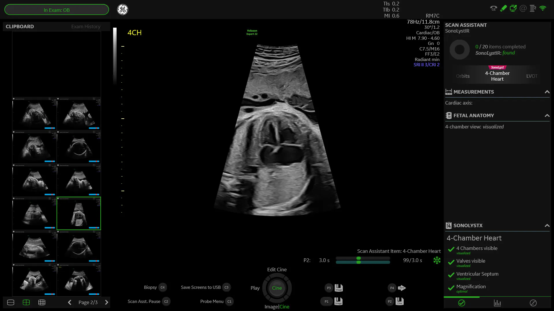
SonoLyst compares the image or view acquired to ensure it meets the accepted clinical standards
SonoLystIR identifies the anatomy visualized and can initiate annotations or measurements
The system streamlines workflow by eliminating the need to freeze and save images during ultrasound screening. Instead, the AI-assisted technology recognizes the fetal anatomy, labels the anatomical part on the screen and captures each image automatically in real time.
Dr. Papageorghiou notes the tool's powerful simplicity during a recent demonstration. "So during the ultrasound scan, I'm scanning over the face and the ultrasound machine's gonna go and grab that face for me," he notes. "I don't need to freeze, it's just going to capture it."
Measurements also are taken automatically, such as with the femur, and saved for review. As each image is acquired, a color-coded signal and checklist appear on the screen, providing information on whether the image is of the highest quality or needs to be retaken.
Intelligent Ultrasound
SonoLystlive continually monitors the completeness of the exam, without changing sonographer workflows. As the sonographer performs the examination, a checklist on the right-hand side of the screen shows the 20 images needed during the screening. As images are obtained, the checklist efficiently marks off the views that have been taken and displays those that remain. Sonographers can still acquire the images in any order, with the SonoLystlive giving clinicians the confidence that nothing has been overlooked during the fetal ultrasound.
Designed as an assistive tool, SonoLystlive provides the flexibility to freeze images and capture them manually, just like with a traditional system. It also provides the versatility to switch back and forth from 2D to 3D during the exam, and from manual to automated.
Dr. Papageorghiou highlights the tool's adaptability, noting, "You can interact with the system as much or as little as you want. So you can just have it running in the background... or you can ask quite a lot of it."
SonoLystlive is advancing ultrasound imaging in prenatal care for greater clinical efficiency and healthier pregnancies.
Interested in viewing Professor Papageorghiou’s lecture? Click here: How AI transforms obstetrical ultrasound.

