
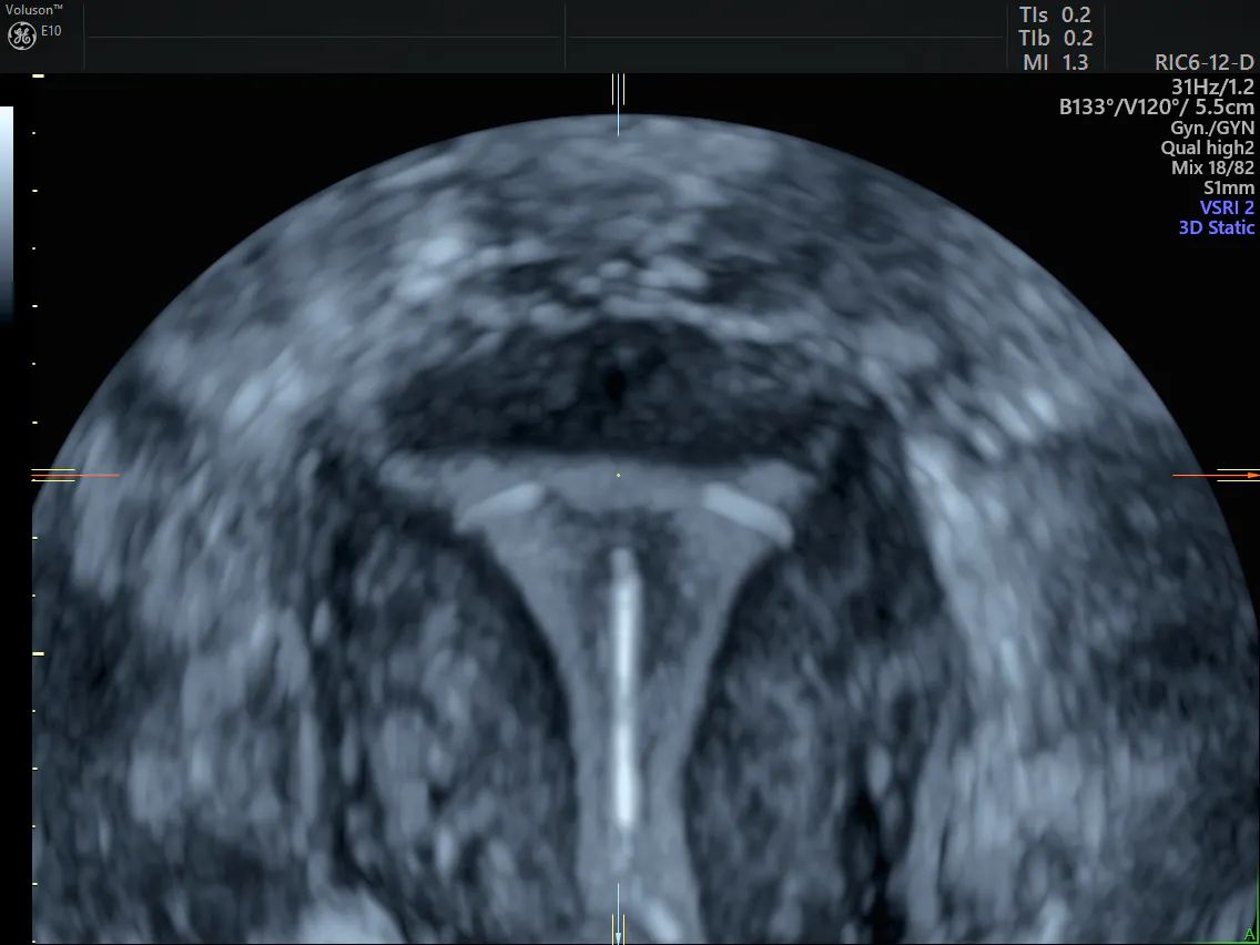
Ultrasound is an easy and accessible screening and diagnostic tool that can be used to assess for uterine and related abnormalities. When it comes to comparing 2D vs 3D ultrasounds, how do these technologies compare? 2D ultrasound delivers a live, real-time view of the pelvis in a radiation-free process that involves high-frequency sound waves. The added benefit of 3D ultrasound is that it adds even more detail to the pelvic ultrasound assessment with the addition of a third imaging plane: the coronal view.
2D vs 3D Ultrasound: Seeing It All With 3D
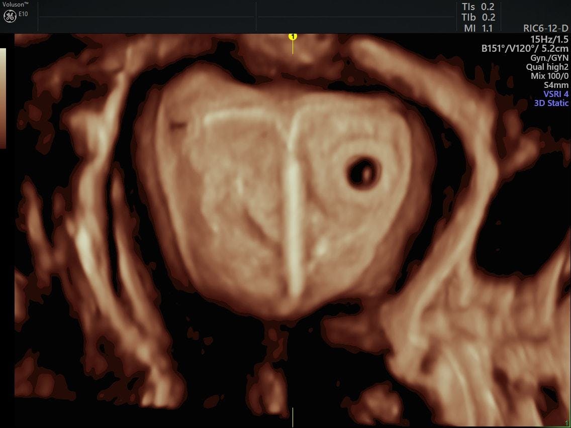
With the coronal plane, the physician can attain a complete view of the gynecological anatomy. It is easy to delineate the fundal contour of the uterus to assess uterine abnormalities as well as the entire inner endometrial lining to thoroughly inspect causes of abnormal bleeding.
Additionally, 3D ultrasound is now the imaging modality of choice for determining Müllerian duct abnormalities of the uterus and locating IUDs within the endometrium or myometrium. It has also been shown to assist significantly with the assessment of intrauterine fibroids and with locating ectopic pregnancies.
3D ultrasound with IUD placement
The Role of Color Doppler
A crucial part of diagnosing pelvic pathology is visualizing the type of blood flow around and within the pathology. Fibroids can have one or many circumferential vessels, polyps can be confirmed by locating their feeder stalk, ectopic pregnancies can be differentiated from cysts by the presence of a "ring of fire," 3D power Doppler can help differentiate between endometrial hyperplasia and carcinoma, and ovarian cysts become more worrisome when an imaging test reveals septations with internal blood flow.
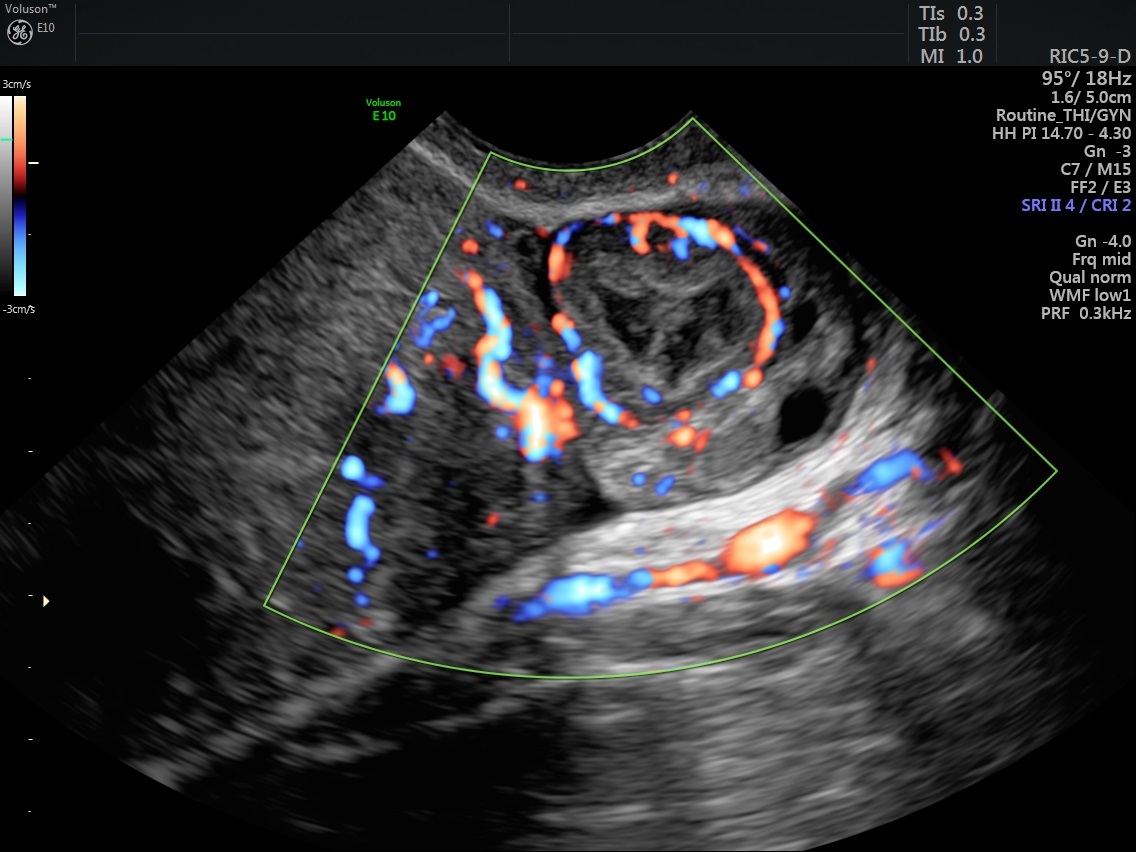
Color Doppler showing flow around corpus lutem
Both color Doppler and HD-Flow™, a bidirectional Doppler that is very sensitive to vasculature, can help physicians make a more informed diagnosis in a gynecologic pathology.
Simplifying Gynecologic Pathology
Despite the complex-sounding disorders that can be encountered in the female pelvis, using ultrasound can be the easiest part of the assessment. Ultrasound technology is more user-friendly, accessible, portable and low-maintenance than ever before.
Due to their portability, affordability and ease of use, ultrasound systems can be used right in the doctor's office. The physician and patient can view results at the time of appointment, which prevents delays that might affect an important diagnosis. This prompt access to test results enables patients to begin treatment immediately, thus improving patient care.
In an increasingly interconnected and interdependent world, collaboration with other physicians and colleagues has become a necessary part of the healthcare process. Image management software integrated with the ultrasound system makes it easy for gynecologists to share test results with the click of a button.
The Future of Gynecologic Imaging
3D ultrasound is the key to the future of effective gynecologic assessment because it enables complete views of the anatomy of the uterus. 2D ultrasound no longer provides all the necessary information for all diagnoses, especially complex and time-sensitive ones. With access to more diagnostic information, physicians can prescribe treatment with confidence and deliver the highest possible quality of patient care.


