An Overview of Key Uterine Abnormalities
Uterine abnormalities appear in roughly 5% of women, with risks that include miscarriage and preterm birth. When a malformation is suspected, 3D ultrasound imaging offers superior diagnostic accuracy, aiding clinical decision-making and patient care.
Defining Uterine Anomalies
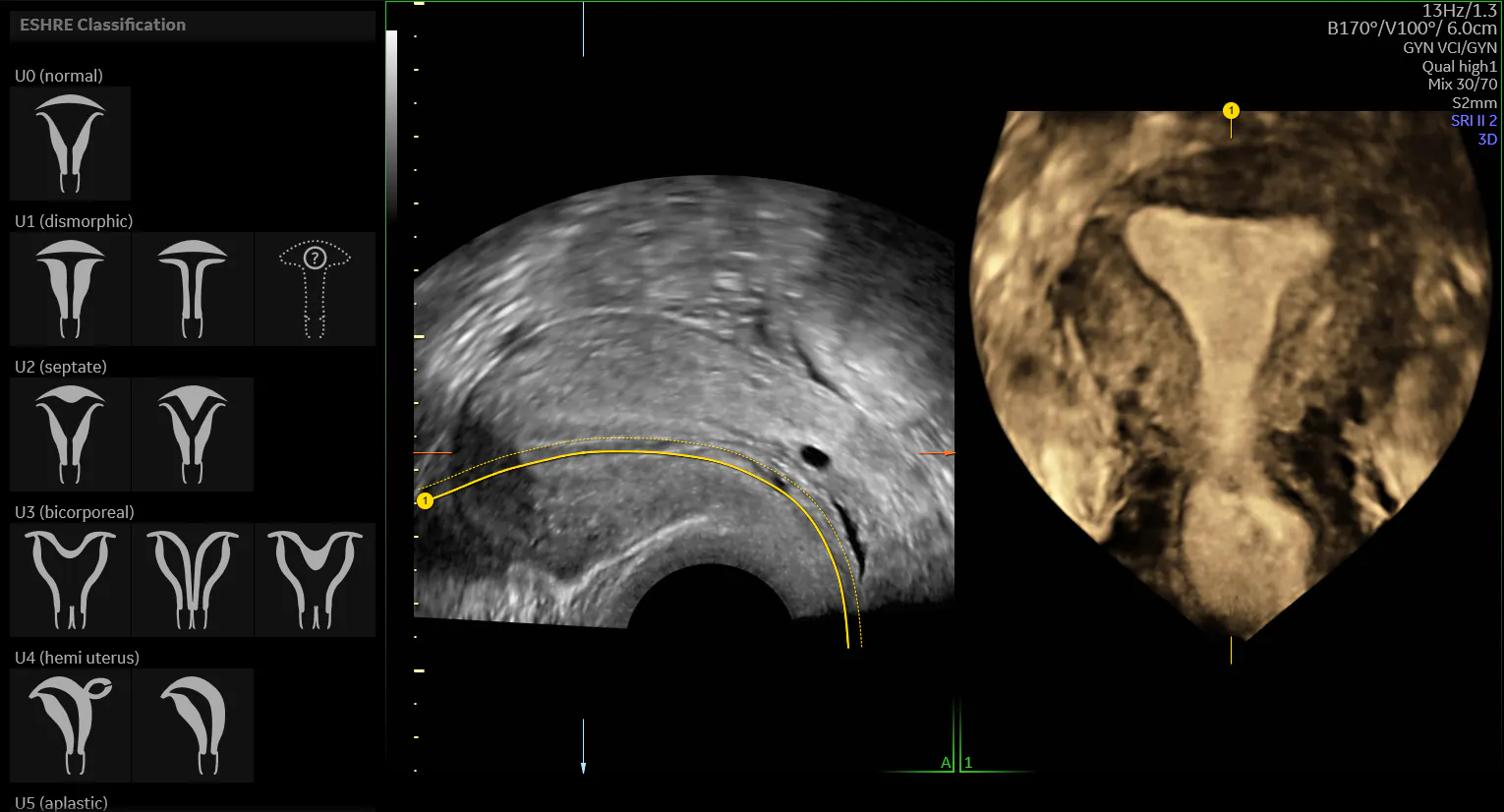
Accurately diagnosing uterine anomalies can be clinically challenging due to varying criteria used to differentiate among the forms of uterine malformation. Amid a lack of consensus, professional societies have developed clinical guidance, such as the classification system from the European Society of Human Reproduction and Embryology (ESHRE) and the European Society for Gynaecological Endoscopy (ESGE). Thus, aided by ultrasonography, clinicians may characterize a normal uterus versus an anomalous one using ESHRE/ESGE criteria.
Based on uterine anatomy, the system outlines six classes of malformations, offering an efficient and straightforward method for identifying and categorizing uterine abnormalities. Per ESHRE/ESGE guidelines, the dysmorphic uterus (U1) appears with a normal uterine outline but a narrow uterine cavity and abnormal internal shape. Similarly, the septate uterus (U2) displays a normal outline but internally has an indentation at the fundal midline greater than 50% of the uterine wall thickness. The indentation may partially or fully divide the uterine cavity.
The bicorporeal uterus (U3) appears abnormal inside and out, with an anomalous fundal outline. An external indentation of more than 50% of the uterine wall thickness is seen at the fundal midline, either partly or fully separating the uterine corpus above the cervix. Classification of the bicorporeal uterus contains three sub-categories, including the most severe form, the bicorporeal septate uterus (U3c). This sub-form includes an absorption defect, with the width of the midline fundal indentation exceeding uterine wall thickness by 150%.
The hemi-uterus (U4) may appear with or without a contralateral rudimentary cavity, while the primary uterine structure is unilateral with a shape resembling a banana. Similarly, the aplastic uterus (U5) may or may not have a rudimentary cavity. Finally, unclassified malformations are designated as U6, per ESHRE and ESGE. The arcuate uterus is considered a normal variant and thus isn't included in the classification.
Separately, the American Society for Reproductive Medicine (ASRM) offers a classification system that's more widely used in the United States, with nine separate anomaly classifications. Among the differences in the two methodologies, the ASRM system groups the normal uterus with the arcuate uterus, which has a subtle concave fundal indentation of less than 1 cm and an angle greater than 90 degrees, per ASRM.
Risk Associated with Uterine Abnormality
Although many women with congenital uterine anomalies remain symptomless, pelvic pain and abnormal bleeding may be warning signs of a malformed uterus. Pregnancy risks associated with uterine abnormality include preterm birth, intrauterine growth restriction, and miscarriage.
[Note: this is from the video]Early research, for example, finds twice the risk of second trimester miscarriage among women with an arcuate uterus. Yet other work suggests that pregnant women with this type of uterus may go on to have live births at nearly the same rate as their "normal" counterparts, suggesting an opportunity for further investigations to truly understand the real-world risk to patients with varying degrees of anomaly.
Ultrasound and Congenital Uterine Anomaly
Three-dimensional transvaginal ultrasound provides a safe, effective method for evaluating the structure of the uterus in patients where a uterine anomaly may be suspected. A pooled analysis in Human Reproduction finds the highest degree of diagnostic accuracy with 3D ultrasound, showing a reported accuracy of 97.6%.
The technique allows for the acquisition of a 3D volume of the uterus, which can be reconstructed and displayed in three intersecting planes, including the critical coronal plane. Given its superior accuracy, 3D ultrasound has become the new standard for detecting abnormalities in the uterine cavity, especially among patients experiencing infertility.
Unlike 2D, 3D offers the advantage of visualizing the external contour. Sonographers can then measure the depth of the septum, view the internal contour and evaluate the depth of the uterine cavity. Once an anomaly has been accurately diagnosed, surgical management may be considered in cases where clinical benefits outweigh risks.
With effective imaging and assessment, clinicians may engage in thoughtful conversations with patients on maternal and prenatal risks and potential treatment avenues to promote the best possible outcome.

