

Congenital uterine anomalies, also called Müllerian duct anomalies, are female reproductive malformations that can increase the risk of infertility or adverse pregnancy outcomes, such as miscarriage, intrauterine growth restrictions and preterm delivery.
According to a study published in Human Reproductive Update, the prevalence of these uterine anomalies is 7.3 percent in the infertile population and 16.7 percent in the repeat miscarriage population. Although uncommon, this condition is treatable. With the right attention and the right kind of imaging and assessments, many hopeful parents can avoid years of painful infertility and repeat miscarriages.
Simplifying Congenital Uterine Anomalies
Different from acquired uterine anomalies that form over time, such as fibroids and endometrial polyps, congenital uterine anomalies are present from birth and occur when the uterus is first forming in the fetus.
The two most common anomalies are the septate and bicornuate uterus. A septate uterus can be partially or completely divided by a muscular or fibrous septum down the center of the uterine body. Due to the limited vascularity of the septum, it is often connected to congenital infertility and repeat early pregnancy loss.
The bicornuate uterus occurs when there is partial fusion failure of the two sides of the uterus, resulting in two separate "horns." This uterus is shaped like a heart with a deep indentation on the fundal surface. The most common symptom is early pregnancy loss and cervical incompetence.
A unicornuate uterus develops only one horn and is just half the size of a normal uterus. There will also only be one fallopian tube connected. There is a significant risk of miscarriage and preterm birth with this anomaly.
A didelphic uterus has two separate uterine bodies, each one with a cervix. It does not cause infertility, but there is a higher risk for preterm delivery, breech delivery and miscarriage due to the limited size of the uterine body.
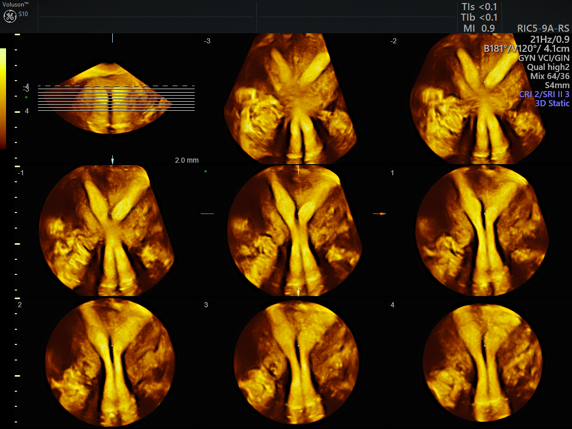
3D ultrasound uterine abnormality
Building Hopeful Futures With Ultrasound
Ultrasound has been used to assess uterine morphology for more than four decades, but in the past, its success rate had varied with congenital uterine anomalies. Other diagnostic methods, such as hysterosalpingography and laparoscopy, both of which are invasive and uncomfortable, and pelvic MRI, which is costly, not as widely available and often requires a significant waiting period, were once necessary to evaluate these anomalies.
Fortunately, the newest form of 3D transvaginal ultrasound can be used to noninvasively and accurately diagnose all congenital uterine anomalies. A study published in the American Society for Reproductive Medicine states that due to the coronal plane displaying both the external contours and internal morphology of the uterus, 3D ultrasound is extremely accurate for the diagnosis and classification of congenital uterine anomalies.
With the current surgical treatments available for many uterine anomalies, pregnancy can generally be attempted three months after surgery, and the prognosis for a successful pregnancy is excellent. This can fast-track family plans for patients who may have worried they would never be able to conceive or carry a pregnancy to term.
The Future of Infertility Assessments
Reproductive failure and adverse pregnancy outcomes are deeply traumatizing to many women, so learning that there is a structural reason for their difficulties can be very reassuring. Knowing that something can be done to increase their chances of achieving and carrying a pregnancy to term gives many women hope and encouragement.
Fortunately, 3D ultrasound offers significant advantages for the assessment of uterine morphology. It is inexpensive, widely available, noninvasive and provides quantitative information on fundal indentation and septum length, which is not possible with other imaging techniques. In addition, 3D ultrasound images are easily shareable with other clinicians and stored ultrasound data can be resliced to provide standardized views of the uterus.

