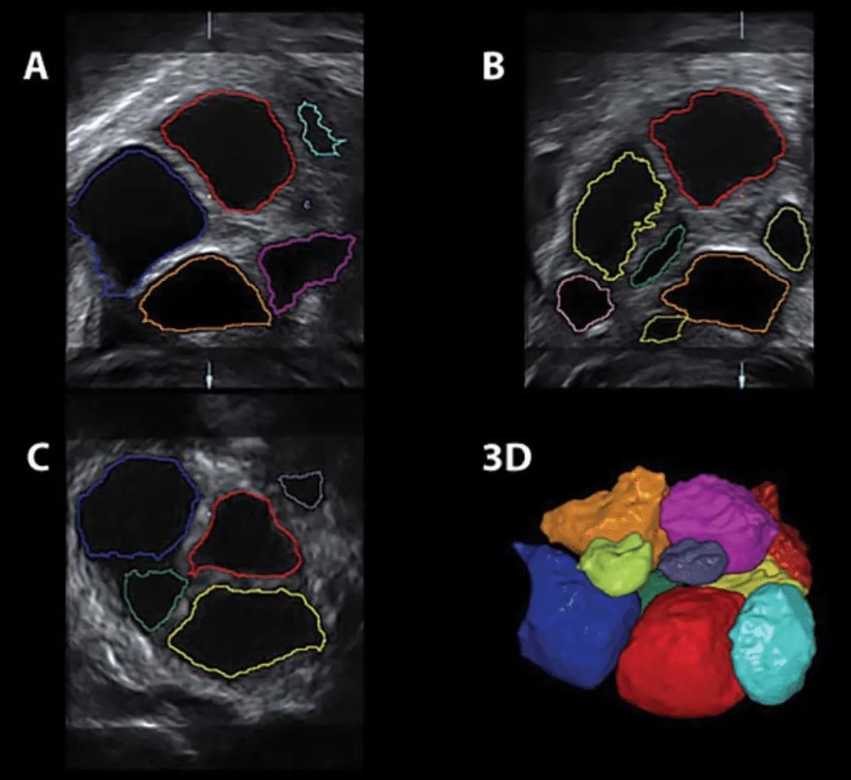
In the past three decades, 2D ultrasound technology has been the gold standard. However, this picture is rapidly changing with the emergence of 3D imaging. Standard 2D ultrasound can visualize horizontal and vertical planes, but 3D ultrasounds can provide a far more accurate image. Translation of a 2D scan to a 3D scan is time-consuming. However, software called sonography-based automated volume calculation (SonoAVC) may change this, and could help improve oocyte retrieval.
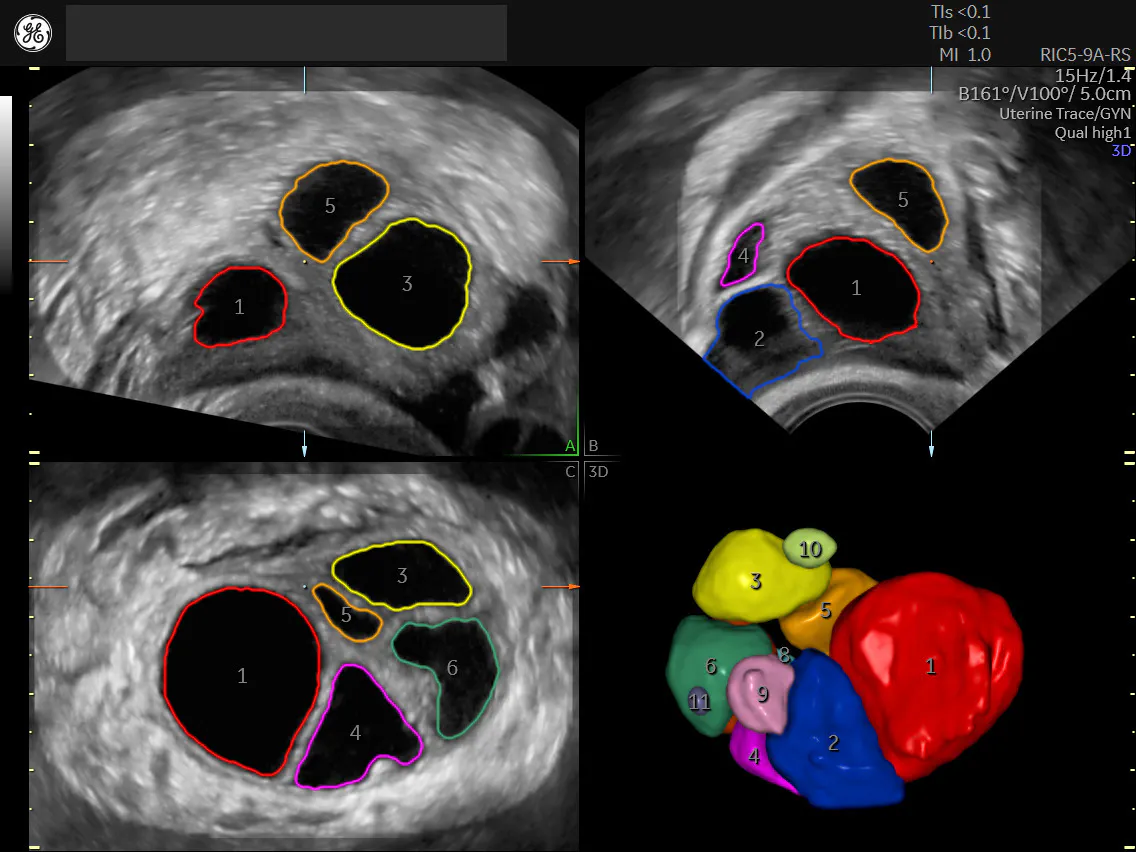
SonoAVC™follicle automatically calculates follicle volume and dimensions
SonoAVC is used for monitoring follicle growth during controlled ovarian hyperstimulation. There is increasing evidence that 3D sonography yields highly reliable follicular volume estimates. The number of small antral follicles is lower when SonoAVC is used to analyze 3D ultrasound data, suggesting that manual assessments may overestimate the number of small follicles. This finding remains to be further explored and confirmed, but reproductive imaging specialists agree that 3D rendering automation is significantly faster compared to measurements with 2D ultrasound imaging.
Are there other benefits to 3D imaging for assisted reproductive technology (ART) as well?
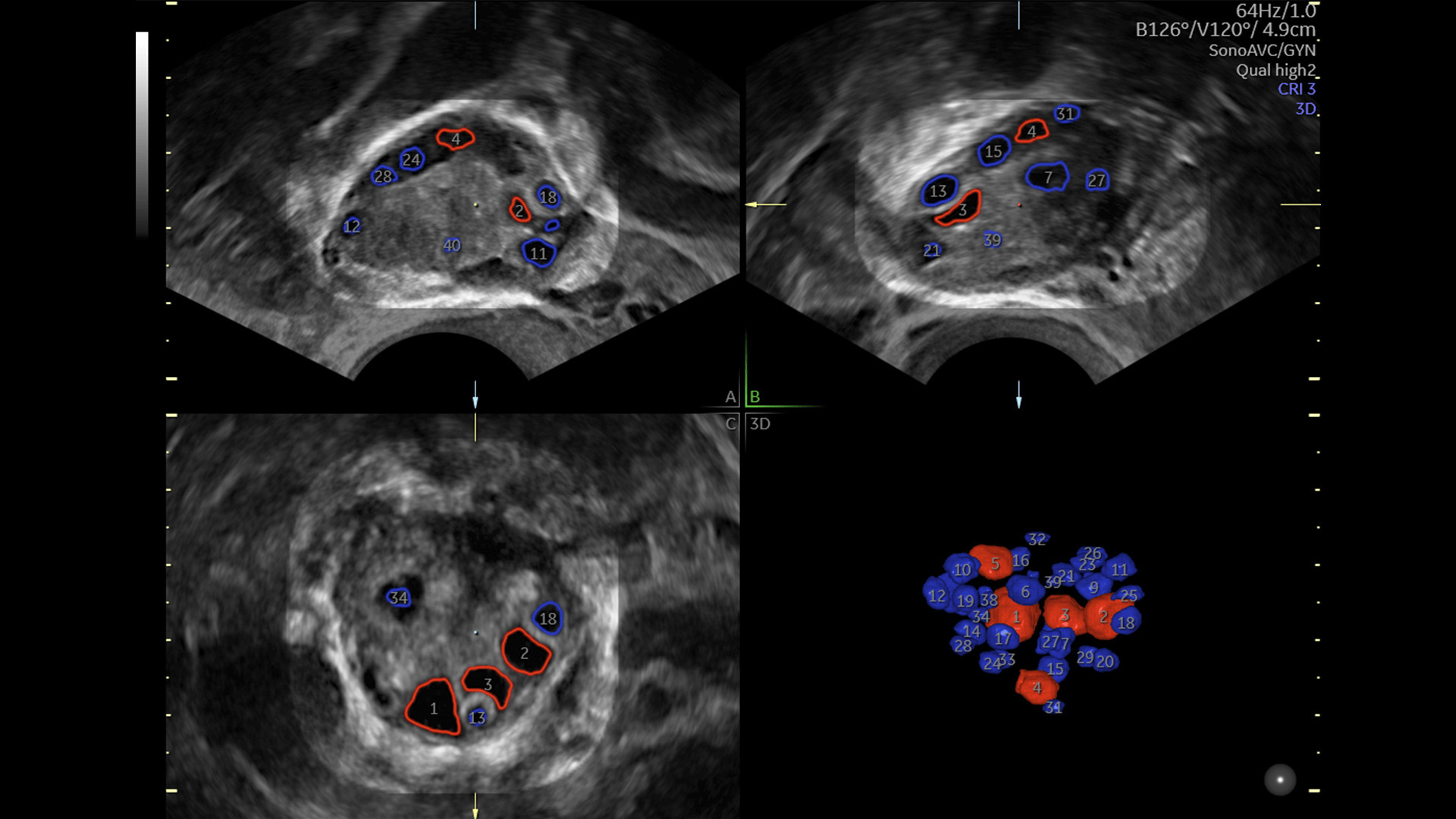
SonoAVC™antral2.0 Sonography-based Automated Volume Count automates ovarian reserve assessment
Assessing 3D Ultrasound With Randomized Clinical Trials
The benefits and efficacy of medical procedures are best established via randomized clinical trials (RCT). Randomization in reproductive medicine is a challenge, due to the complex nature of the technologies involved and the many steps required to assure minimal bias. Blinding — a basic tenet of RCTs — may also be a problem, as investigators attempt to convince patients to participate while explaining that the allocation of their case to the control or intervention arms of the study cannot be disclosed to them.
The first randomized trial to investigate the benefit of 3D ultrasound was performed in a group of 40 patients whose partners were diagnosed with male factor infertility. Participants were randomized into a control group with either standard 2D ultrasound, or the study group for whom 3D SonoAVC with automated follicle volume count was applied. All outcome parameters — including the level of oocyte retrieval and pregnancy rate — were similar, but more mature oocytes were fertilized in the 3D group. A second 3D-automated follicular volume trial was presented in 2017. This randomized trial compared automated 3D with 2D manual methods of follicle tracking and assessed ART outcome in 130 patients by studying the number of mature oocytes retrieved, fertilization, pregnancy and live birth.
Use of automated 3D monitoring added no advantage over manual 2D ultrasound tracking in improving clinical or laboratory outcomes during IVF cycles. The former significantly reduced monitoring time, however, and this was considered a potential time- and cost-saving benefit. However, because patient selection criteria in the two studies were not the same, comparing these results or drawing firm conclusions from their findings is difficult.
Saving Time With 3D Ultrasound
Time-saving practices during follicular monitoring for ART and oocyte retrieval are an important practical consideration. Patient experience may be enhanced and costs reduced. Also, due to computerized modelling, data may be compiled and different responses to drugs categorized to optimize the trigger for egg-retrieval timing.
Based on the capabilities of 3D ultrasound technologies, it is perhaps safe to say that other clinical advantages are likely to emerge over time. They are difficult to demonstrate readily, however, due to more than 200 potential confounders of outcomes following IVF. Only larger trials using strict patient selection criteria and appropriate blinding may demonstrate the effect on fertilization or other key outcomes.
Learn more about the benefits of SonoAVC™ in the Assisted Reproductive Setting from Prof. Palumbo et al here.