

Adenomyosis is a common gynecological condition that can cause heavy, prolonged periods and chronic pelvic pain. The true incidence is unknown, and the prevalence varies widely from 5 to 70 percent.1 And just like endometriosis, adenomyosis often goes undetected.
Diagnosing Adenomyosis with Ultrasound
The Advantage of Ultrasound in Detecting Adenomyosis
A Q&A with Dr. Lisbet Hanson on ultrasound findings which can indicate adenomyosis and how ultrasound can lead to a faster diagnosis.

Adenomyosis is a common gynecological condition that can cause heavy, prolonged periods and chronic pelvic pain. The true incidence is unknown, and the prevalence varies widely from 5 to 70 percent.1 And just like endometriosis, adenomyosis often goes undetected.
Lisbet Hanson, M.D., Medical Director of Sentara’s Women’s Health Program at Virginia Beach General Hospital, shares ultrasound findings which can indicate adenomyosis and how ultrasound can lead to a faster diagnosis.
Q: Countless women experience severe menstrual bleeding and excruciating cramps, but adenomyosis is one diagnosis that is often overlooked. How can ultrasound play a role in changing that?
A:First, I think we need to educate our patients more about adenomyosis and endometriosis. As clinicians, if a woman says she’s having horrible, debilitating periods, we need to figure out what’s going on. I think ultrasound is an incredible tool to help make a diagnosis quickly. In the past, adenomyosis was typically diagnosed in older women after a hysterectomy. But with ultrasound, we are now seeing it in younger women as well—and clinicians are making the diagnosis in the office long before a surgical procedure. We are then able to start treatment with an IUD, oral contraceptives or progesterone.
Q: It’s estimated that transvaginal ultrasound has a sensitivity of 79 percent and specificity of 85 percent for the detection of adenomyosis.10 What are some of the common ultrasound findings in patients with adenomyosis?
A:When you look at the ultrasound images, you can see patterns that could indicate adenomyosis. An irregular or interrupted Junctional Zone should raise concern. You may notice an enlarged or asymmetric uterus (one wall is bigger than the other). Another indication is linear shadowing in the wall of the uterus without the presence of a fibroid. It’s also common to see bright lines or stripes that look like streaks of light. There is a consensus statement from the Morphological Uterus Sonographic Assessment group on the ultrasound criteria for adenomyosis.2
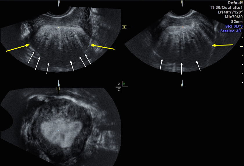
Adenomyosis showing asymmetric uterus (yellow arrows) and linear shadowing (white arrows)
Q: How can color Doppler enhance your ability to diagnose adenomyosis and why is this feature so important?
A: Color Doppler is also helpful in separating adenomyosis from fibroids. You can look at the blood vessels in the wall of the uterus and see the blood flow has very different patterns. For adenomyosis, the vessels run translesionally, rather than in a circular pattern with myomas. It’s an important distinction because adenomyosis doesn’t have a pseudocapsule—making surgical excision incomplete and very difficult in most cases.
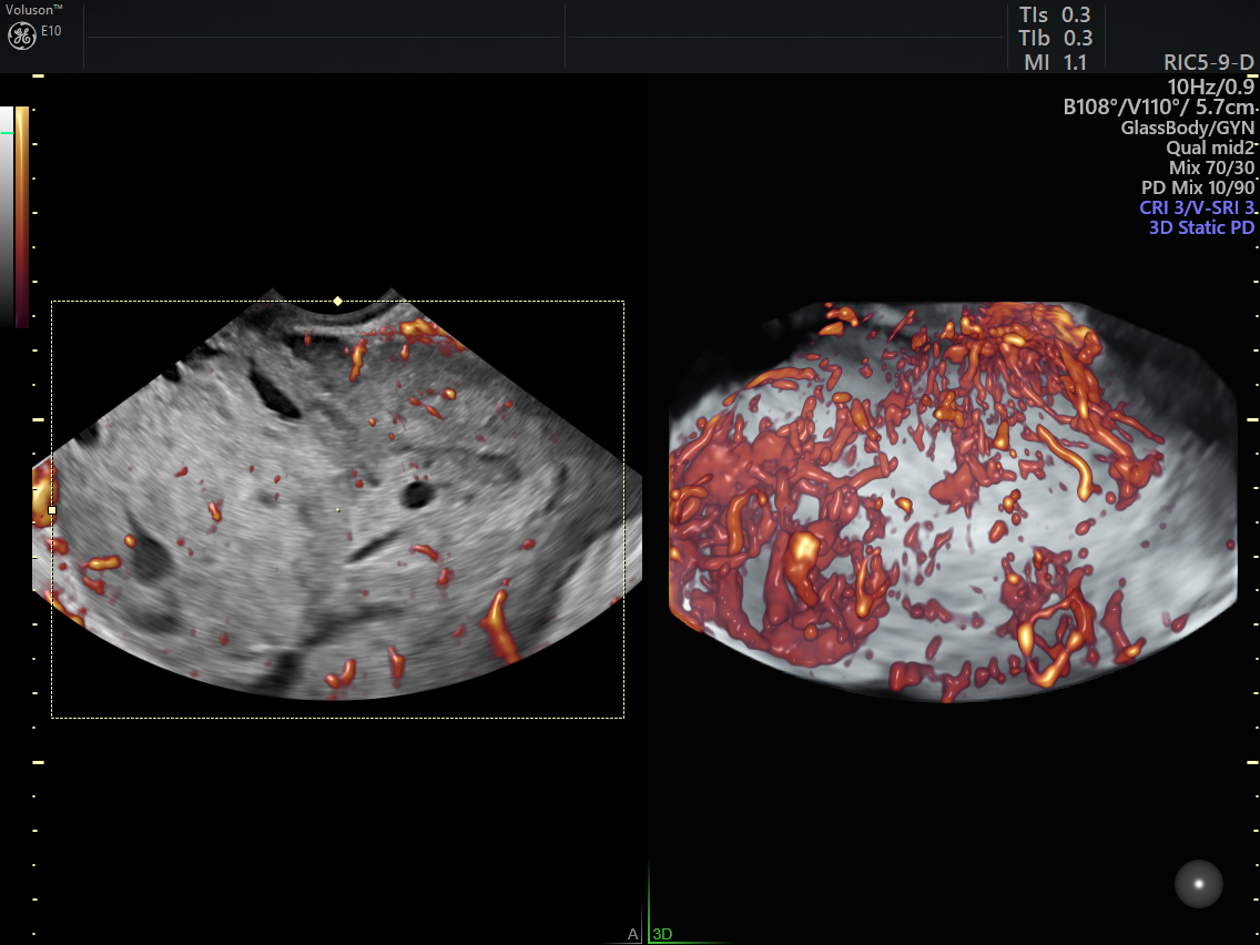
Adenomyosis vascular pattern shows blood flow through the area, in a non-circular pattern
Q: How long have you been using Voluson™ ultrasound by GE Healthcare and how does it help in your daily practice?
A:I started using Voluson LOGIQ Book XP about 18 years ago. It was a great little tool, the size of a laptop. Lightweight and small, I could roll it from one exam room to the next without worrying about running over cables or bumping into door frames and injuring the probe. In residency, I had quite a bit of exposure to obstetrical ultrasound, but very little GYN. When I started doing more and more GYN care, I realized I would have to learn how to do GYN ultrasounds, interpret the images and write reports. I went to several courses and asked a friendly sonographer friend if she would mind observing me and giving me some tips. Pretty soon I became very comfortable with the basic GYN ultrasound exam. I built on it from there. As the research and technology improved and discovered better ways to look at the uterus, the myometrium, the endometrium, and the junctional zone I started doing 3D ultrasound on every GYN patient. We now use a Voluson E6 in the office and it has its very own room, rather than being wheeled from one exam room to the next. The 3D gives an unparalleled view of the fundus and Junctional Zone. The options for image animation and manipulation make it easier and easier to see things like adenomyosis that were once only diagnosed by the pathologist at the time of hysterectomy.
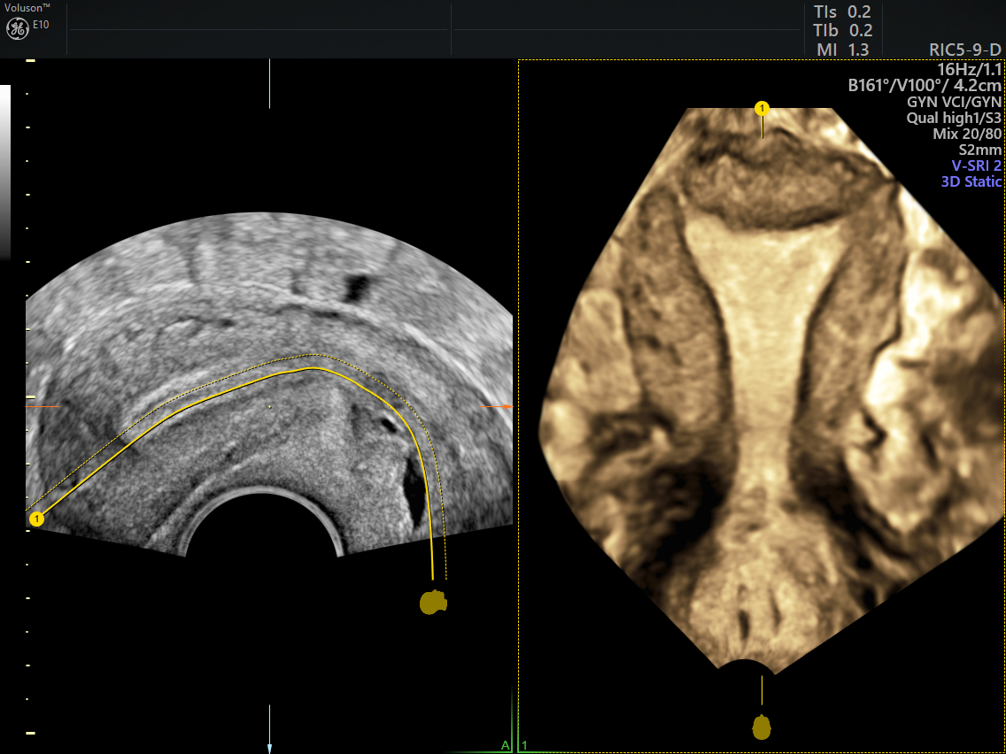
3D OmniView of uterus with a normal junctional zone between the endometrium and myometrium
Q: How important is it for physicians to be able to perform ultrasounds and stay current with the related research and advancements?
A:The technology is changing quickly, and you have to keep up. That’s the excitement of medicine really, right? We are constantly challenged to learn new information. I think you should feel comfortable doing ultrasounds and understanding the capabilities and limitations. The more experience you get doing ultrasounds, the better you get at performing ultrasounds and interpreting the images. It’s extremely important for gynecologists and patients.
-
Transvaginal ultrasound is gaining ground as a noninvasive and low-cost way to obtain an adenomyosis diagnosis for patients of all ages.
-
What is ultrasound used for? Ultrasound has many applications for women's health, far beyond just obstetrics. Read our collection of FAQs.
-
Although not cancer, adenomyosis can cause severe symptoms that negatively impact a woman's quality of life.
-
Providers should follow the latest AIUM ultrasound guidelines to ensure they're equipped to provide high-quality, up-to-date care to their patients.
-
Try out this fun exercise and learn more about the benefits of ultrasound in GYN diagnosis.
-
Pelvic pain can be multifactorial in origin – the single most common cause is endometriosis. Transvaginal ultrasound has been shown to detect endometriosis with high accuracy.
-
Learn more about the 4-step ultrasound evaluation as suggested by the International Deep Endometriosis Analysis (IDEA) group.
-
Endometriosis has complex symptoms that can mimic other medical conditions, contributing to the delay in diagnosis. And there’s the common misconception that periods equal pain. Many women simply accept the burden and don’t bother seeking help, while countless others are dismissed by doctors for exaggerating pain.
Li J, Chung J, Wang S, Li T, Duan H, The Investigation and Management of Adenomyosis in Women Who Wish to Improve or Preserve Fertility, Biomed Res Int. 2018 Mar 15
Van den Bosch T, de Bruiin AM, de Leeuw RA, Dueholm M, Exacousos C, Valentin L, Bourne T, Timmerman D, Huirne JAF, A sonographic classification and reporting system for diagnosing adenomyosis, Ultrasound Obstet Gynecol. 2018 May 22
Maheshwari, A.; Gurunath, S.; Fatima, F.; Bhattacharya, S. (2012). "Adenomyosis and subfertility: A systematic review of prevalence, diagnosis, treatment and fertility outcomes". Human Reproduction Update. 18 (4): 374–392.

