

Adenomyosis is a relatively common, benign uterine condition. The tricky part of treating adenomyosis is that its symptoms — pelvic pain and heavy or painful menstruation — often mimic those of diseases such as endometriosis, making adenomyosis difficult to diagnose.
Magnetic resonance imaging (MRI) was previously considered the best way to establish an adenomyosis diagnosis, but ultrasound is steadily becoming the preferred imaging tool thanks to its high degree of accuracy and ability to inform timely, effective treatment.
Making a Diagnosis: Adenomyosis Ultrasound or MRI
Transvaginal ultrasound is now superseding MRI for diagnosing adenomyosis in many practices due to its wide availability, low cost and quick exam time.
Research has illustrated that the accuracy of ultrasound is comparable to that of a uterine adenomyosis MRI. One study in Obstetrics and Gynaecology reports that transvaginal ultrasound has a sensitivity of 80.8 percent and a specificity of 61.4 percent when it comes to detecting adenomyosis. According to the American Journal of Roentgenology, MRI operates with a specificity of 96 percent and a sensitivity of 63 percent.
Researcher Caterina Exacoustos, MD, is an international authority on adenomyosis, endometriosis, transvaginal ultrasound and infertility. In an editorial she co-wrote for Fertility and Sterility, Exacoustos spoke to the strengths of ultrasound for adenomyosis diagnosis: "Diagnosing adenomyosis should now be based mostly on endovaginal ultrasound since it is an accurate and easy method [that] can be performed on all types of patients."
But how can clinicians identify adenomyosis using ultrasound?
What Does an Adenomyosis Ultrasound Look Like?
Adenomyosis occurs when ectopic endometrium invades the myometrium and leads to hyperplasia and hypertrophy of the surrounding smooth muscle. While the condition is classified as benign, it can become malignant.
A sonographer's first clue to adenomyosis is often an ill-defined lesion within the myometrium. A lesion may have an irregular contour, no rim, no edge shadows or a fan-shaped shadow.
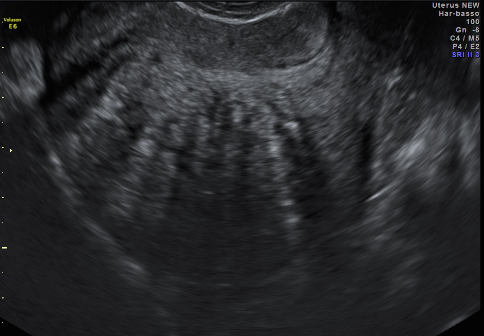
Diffuse adenomyosis, classic fan-shaped shadowing on adenomyosis ultrasound
Diffuse adenomyosis, classic fan-shaped shadowing on adenomyosis ultrasound
Other indicators on adenomyosis ultrasound images may include:
- Myometrial cysts
- Hyperechoic islands
- Subendometrial lines and buds
- Translesional flow
- A thickened, irregular or ill-defined junctional zone
- Myometrial anterior-posterior asymmetry
- An enlarged uterus
While all these findings may indicate adenomyosis, there are three key signs that are the most important for clinicians to identify:
- Ectopic endometrial glands: These manifest as echogenic nodules and striations radiating from the endometrium into the myometrium. When these glands contain fluid, myometrial cysts and fluid-filled striations may also be visible.
- Muscular hyperplasia and hypertrophy: This appears as globular uterine enlargement and asymmetric myometrial thickening, often with thin "venetian blind" or streaky shadowing.
- Hypervascularity: Adenomyosis increases uterine vascularity, which shows up on color Doppler as a pattern of penetrating vessels. It is important to distinguish these from peripheral vessels, which indicate the presence of fibroids.
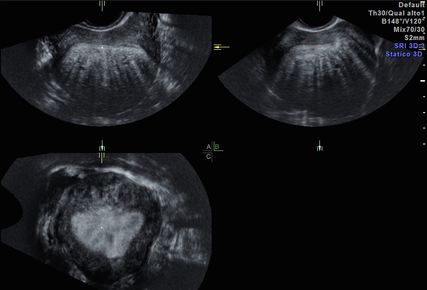
Using 3D ultrasound also allows a clinician to obtain cine clips and coronal images that provide an overview of the entire endometrial-myometrial border. This broad view is crucial given that adenomyosis can present in either a focal or diffuse form.
The Changing Face of Ultrasound
As ultrasound becomes more and more popular for diagnosing adenomyosis, researchers are working to streamline the diagnostic parameters. Ultrasound in Obstetrics & Gynecology recommends a specific classification and reporting system for adenomyosis based on the Morphological Uterus Sonographic Assessment (MUSA) criteria. Similar to how the International Ovarian Tumor Analysis guidelines aim to classify adnexal masses, the MUSA criteria offers doctors concrete parameters for describing adenomyosis, such as lesion size and location in the uterus.
Ultrasound offers a significant benefit not only for clinicians, but also for patients who are coping with adenomyosis. With effective diagnostic tools, we can work toward more efficient treatment and a better understanding of this disease overall.