

Retained products of conception (RPOC) are estimated to complicate between 1 and 19% of pregnancies—and are probably more common after miscarriage or termination of pregnancy.1 Without a timely and accurate diagnosis, women can face potentially harmful and unnecessary surgery, infertility, and even death.
Postpartum Ultrasound and the Importance of Scanning for RPOC
Help Patients Avoid Unnecessary Interventions
Learn how Voluson™ ultrasound technology can provide critical clinical information that could help avoid devastating complications when scanning for RPOC.

Retained products of conception (RPOC) are estimated to complicate between 1 and 19% of pregnancies—and are probably more common after miscarriage or termination of pregnancy.1 Without a timely and accurate diagnosis, women can face potentially harmful and unnecessary surgery, infertility, and even death.
Excessive bleeding, pain, and fever are the first clues that your patient may have retained products of conception—but making the diagnosis can be a challenge. With no clearly defined diagnostic criteria, evidence-based guidelines, or treatment protocols to follow—your patient is depending on you and your technology for answers.
That’s why experts, including Dr. Larry Platt of the Center of Fetal Medicine and Women’s Ultrasound in Los Angeles, follow their own methodology when they suspect a patient may have remaining intrauterine tissue after a miscarriage, termination of pregnancy, delivery or caesarian section.
“There’s no question that you have to worry about this complication, and it’s ultrasound first,” Platt stressed.
Voluson™ ultrasound technology can provide critical clinical information that could help avoid devastating complications. It’s estimated that nearly 20% of RPOC’s have increased vascularity and in such cases simple D&C may lead to massive hemorrhage.2,3 The findings could spare patients from unnecessary surgery, and prevent possible infection, intrauterine adhesions, and scarring—all of which are linked to infertility and recurrent pregnancy loss.4
“When you suspect RPOC, do you want to rush to go do a D&C? The risk of perforation and the risk of complications are much greater. By doing these diagnostic tests you can reduce the risk to the patient and avoid a therapeutic intervention that may not be needed. Maybe you just give her some type of Ergot derivatives to make her uterus contract without doing a surgical procedure.” Platt said.
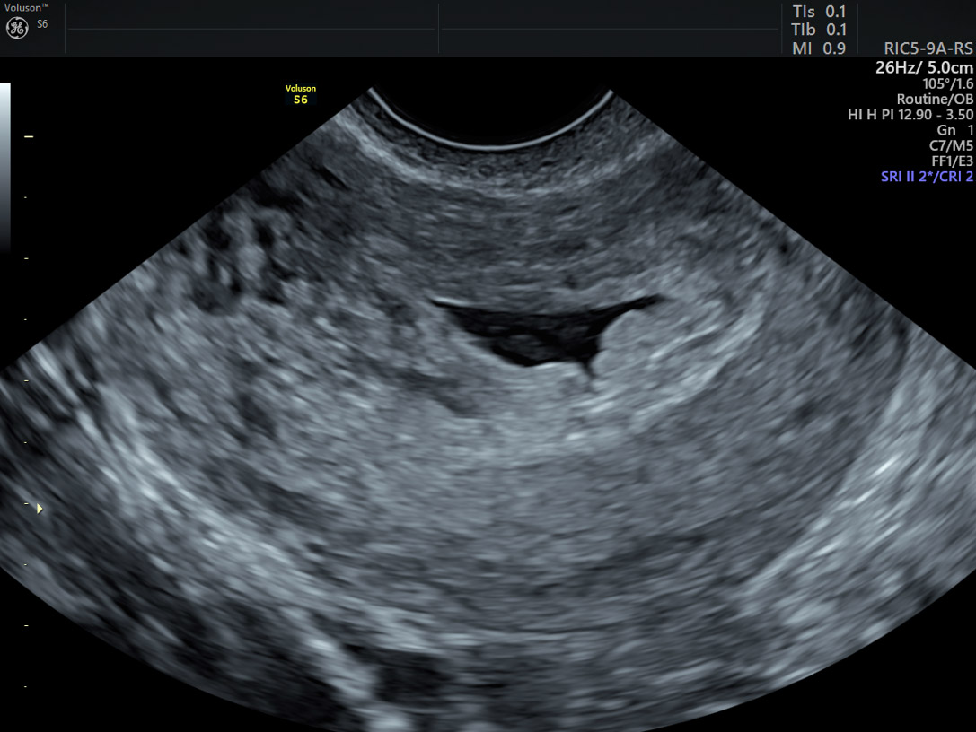
Advanced 2D and 3D ultrasound provide stunning high-resolution views of the uterine cavity—allowing physicians to evaluate the thickness of the endometrial lining and help detect masses and echogenic fluid that could confirm RPOC.5
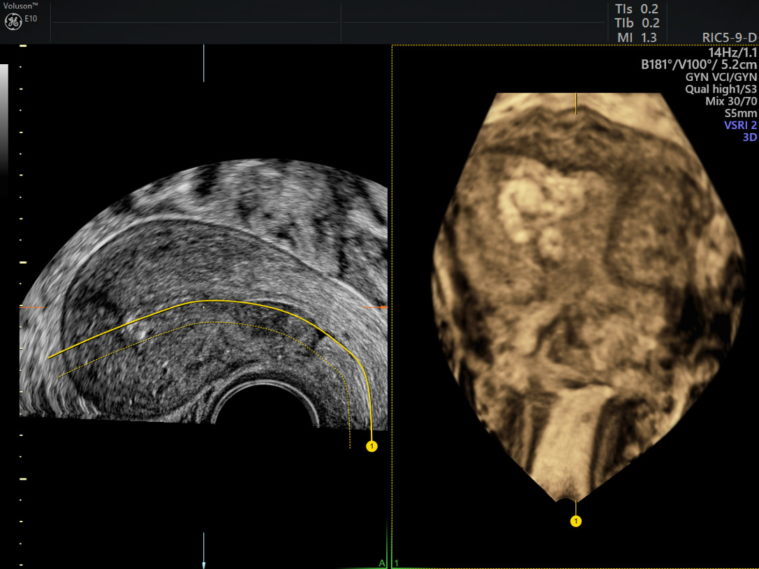
“I favor 3D because it’s the only way to see the C-plane and get a good look at the lining of the uterus when you cut it down the center. You can see the thickness of the endometrial lining, which you can expect to be thinning in the days and weeks after delivery. If you see large echoes filling the endometrial cavity—not just blood—you know there’s a problem,” Platt explained.
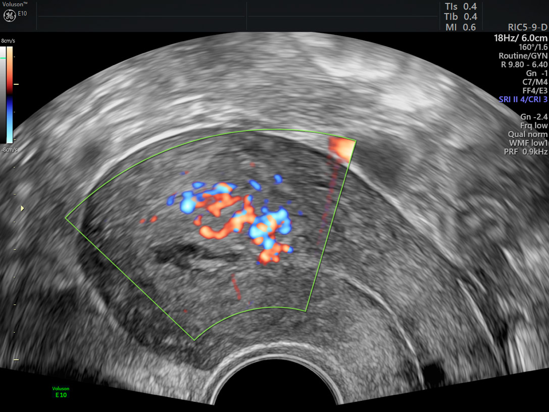
Color Doppler is also a valuable tool in detecting any remaining placenta tissue because it can show evidence of vascularity. Voluson’s Radiantflow has increased sensitivity, allowing for easy, fast visualization of blood flow, even in the tiniest of vessels. Another feature, SlowflowHD, improves your ability to visualize blood perfusion by expanding the range of visible blood flow to include low-velocities.
Platt says these types of innovations have enhanced his capabilities to make crucial diagnoses in gynecology—and the technology is improving all the time. He’s quick to add that GE Healthcare makes it easy for physicians to keep up.
“The longevity of the equipment is enhanced tremendously by software upgrades. With Voluson, you can buy something now and it won’t be outdated in a year. The platform plays a vital role in creating cost-effective medicine—and your ability to stay ahead of the game and stay in the game!”
-
With VolusonTM ultrasound systems you can easily visualize anatomy in the coronal plane with a 3D volume and gain insights to help detect and diagnose gynecological conditions earlier than ever before. Learn more about the extraordinary GYN imaging and analysis tools Voluson offers to help support the accuracy of your exams.
-
What is ultrasound used for? Ultrasound has many applications for women's health, far beyond just obstetrics. Read a collection of frequently asked questions to learn more about some of the uses for ultrasound in your gynecology practice.
-
The ability to make a fast, accurate diagnosis is one of the many benefits of 3D ultrasound technology. Read on to find our fill list of benefits.
-
Find out why ultrasound exams right in the office can lead to more accurate care, time-savings for patients, and lower costs for the practice.
-
Learn more about how a pelvic ultrasound during routine Well-Women exams can improve a clinician's ability to accurately diagnose gynecological concerns.
-
Learn how to image for pelvic floor dysfunction in this educational video.
Hamerlynck, Tjalina W.O. et al. An Alternative Approach for Removal of Placental Remnants: Hysteroscopic Morcellation. Journal of Minimally Invasive Gynecology, 2013, Volume 20, Issue 6, 796 – 802.
Kamaya, A., Petrovitch, I., Chen, B., Frederick, C. E. and Jeffrey, R. B., Retained Products of Conception. Journal of Ultrasound in Medicine, 2009;28: 1031-1041.
Aseeja V. Management of retained products of conception with marked vascularity. J Turk Ger Gynecol Assoc. 2012;13(3):212-4.
Yu D., Wong Y.-M., Cheong Y., Xia E., Li T.-C. Asherman syndrome-one century later. Fertility and Sterility, 2008;89 (4), pp. 759-779.
Sellmyer MA, Desser TS, Maturen KE et al, Physiologic, histologic, and imaging features of retained products of conception. RadioGraphics, 2013;33:781–796.
Hooker, Angelo B. et al., Long-term complications and reproductive outcome after the management of retained products of conception: a systematic review. Fertility and Sterility, 2016; Volume 105, Issue 1, 156 - 164.e2.

