

Since their invention in the late 1950s, ultrasound systems have offered clinicians a noninvasive way to visualize the inside of the body. Standard 2D ultrasonography aids gynecologists by using high-frequency sound waves to create images of the pelvic anatomy, including the uterus, ovaries and surrounding tissue.
But the benefits of 3D ultrasound for your practice are even greater. This technology promises to advance patient care by improving the gynecologist's ability to make confident and informed decisions.
2D Versus 3D Ultrasound
You're familiar with traditional ultrasound, which provides flat, 2D images from just the sagittal and transverse planes of the patient's body. Recent innovations in technology are now able to reconstruct the coronal plane using 3D ultrasound images that more realistically represent the internal organs.
Real Clinical Benefits of Adding 3D Ultrasound
Even if you've never used 3D ultrasound yourself, you may recognize it from its use in obstetrics. This technology is responsible for providing physicians and expectant parents with fetal images that are far more lifelike than conventional sonograms.
Yet the benefits of 3D ultrasound extend well beyond pregnancy. Indeed, a growing body of evidence suggests that this approach can be a useful advancement from standard ultrasound and, in many cases, improve the physician's ability to accurately diagnose certain gynecological concerns. The benefits of adding 3D ultrasound include:
- Diagnosing congenital anomalies. Nearly 17 percent of women prone to recurrent miscarriages in one study had congenital uterine anomalies. Compared with traditional transvaginal ultrasonography — which identifies about 60 to 82 percent of these anomalies — 3D ultrasound can diagnose them with 88 to 100 percent accuracy, according to research published in the Journal of Ultrasound in Medicine.

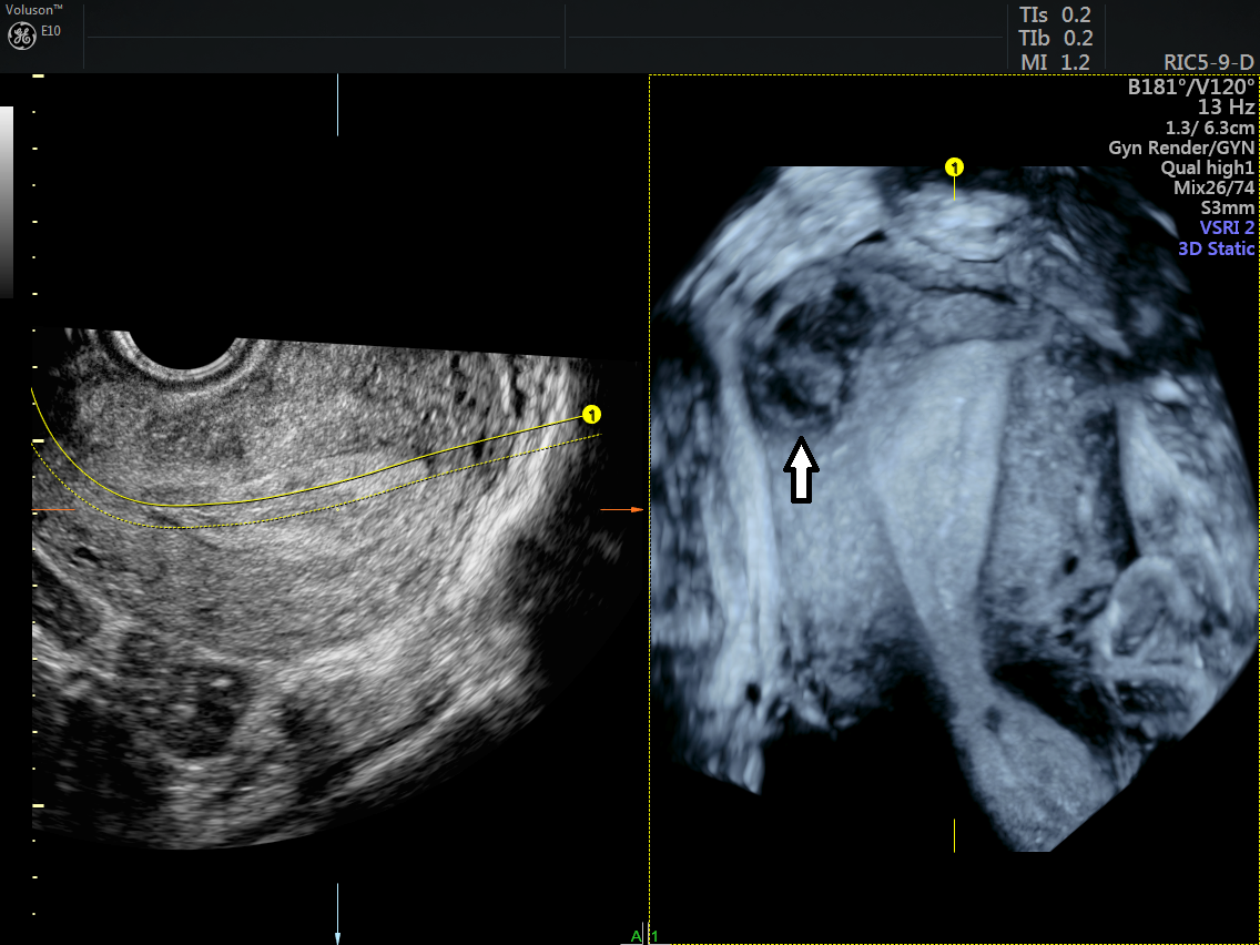
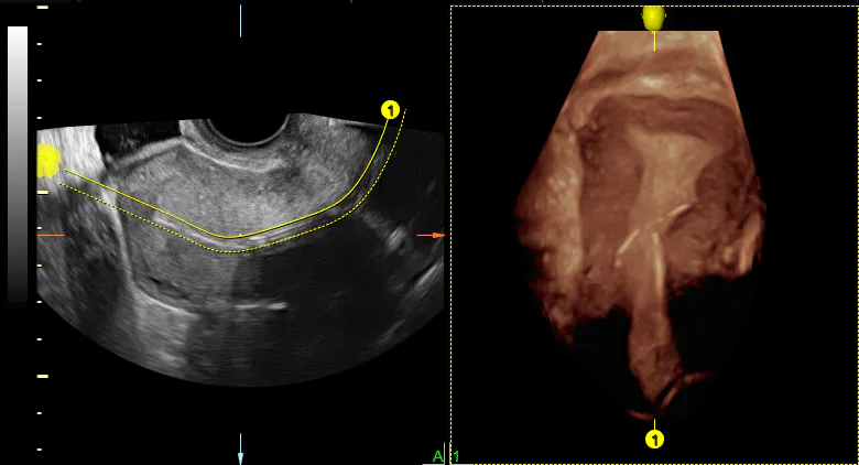
3D imaging using saline infusion to demostrate the uterine cavity of a septate uterus
- Locating fibroids and polyps. Research published in Ultrasonography suggests that 3D ultrasound is more useful than 2D technology at pinpointing the location of fibroid tumors and endometrial polyps in some patients.
3D image of the uterus with a cornual fibroid
- Identifying uterine adhesions. Also known as uterine synechiae, uterine adhesions can impair fertility. 3D ultrasound appears to be more effective than traditional procedures such as a hysterosalpingogram at identifying these adhesions.
- Examining intrauterine device (IUD) placement. Gynecologists typically use 2D ultrasound to place IUDs, but this approach doesn't completely visualize the full IUD. By using 3D ultrasound, physicians can see the entire device in the coronal plane while keeping exam times short. This has also been shown to help improve the detection rate of IUDs that have embedded into the uterine tissue, another study published in Ultrasonography noted, identifying a significant source of pain and abnormal bleeding in some patients.
3D ultrasound image demonstrating a malplaced IUD
Immediate, Accurate Results
Given all the benefits of 3D ultrasound, the technology significantly improves a gynecologist's ability to make accurate diagnoses. Researchers from Massachusetts General Hospital and Harvard Medical School found that 3D ultrasound helped provide gynecologists with additional information in 53 percent of patients who had abnormal findings on a standard, 2D ultrasound. That means you can feel confident making immediate clinical decisions — and when 3D technology is at your fingertips, your patients don't even need to leave your office for advanced imaging.


