

It’s estimated that 80% of women will have fibroids by the time they turn 50.1-4 And the risk is higher for black women—who are three times more likely to develop uterine leiomyomas than white women. Detection and accurate diagnosis are critical because fibroids could contribute to infertility and prompt you to explore the possibility of cancer.
Detecting Fibroids with Ultrasound
More Certainty in Finding and Monitoring Uterine Leiomyomas
Discover how Voluson’s biggest innovations, Radiantflow™, helps distinguish polyps from fibroids and leads to a more confident diagnosis.

It’s estimated that 80% of women will have fibroids by the time they turn 50.1-4 And the risk is higher for black women—who are three times more likely to develop uterine leiomyomas than white women. Detection and accurate diagnosis are critical because fibroids could contribute to infertility and prompt you to explore the possibility of cancer.
Watch and wait. That’s the strategy for Paulien, who has been living with fibroids for more than two decades. The thought of having uterine tumors would send most women into a panic, but 53-year-old Paulien, from Amsterdam, is convinced they are harmless fibroids—not life-threatening cancer. And a lot of her confidence comes from ultrasound.
“You have to go by your doctor’s experience and what he sees on ultrasound. Is it a fibroid or are there signs of something else? I trust my doctors and I trust ultrasound,” Paulien said.
Paulien isn’t your typical patient. She’s also a general medical sonographer at a Medical Treatment Center. She had seen uterine leiomyomas on other patient scans before her initial diagnosis, but she didn’t know the clinical implications of fibroids or treatment options because she had limited experience in gynecology.
“I was in my early 30s and my gynecologist found one small fibroid while changing my IUD. I wasn’t really concerned. I didn’t have any symptoms like pain or heavy bleeding, so we decided to monitor the fibroid regularly with ultrasound,” Paulien said.
According to Lawrence D. Platt, M.D. of the Center of Fetal Medicine and Women’s Ultrasound in Los Angeles, it can be difficult to detect leiomyomas with a pelvic exam. To him, it’s about having the right tools. And diagnosing fibroids is especially important when a patient is struggling with infertility.
“Without ultrasound, you can miss a significant number of fibroids that are one, two, or three centimeters. Small fibroids can get in the way of the endometrial cavity and can contribute to infertility. So, if a woman can’t get pregnant—those are the things we look for,” Platt explained.
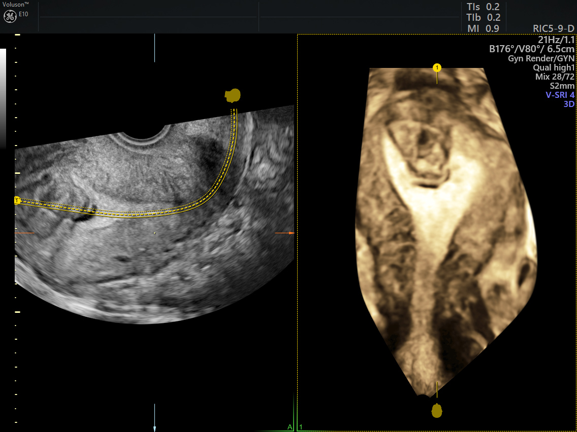
Uterine fibroid invading the endometrial cavity
Dr. Platt relies on Voluson™ ultrasound systems from GE Healthcare for a more complete evaluation of the uterine cavity. The gynecological expert appreciates the flexibility of having so many versatile features in one machine—emphasizing the importance of 3D technology. In some cases of suspected fibroids, Dr. Platt will perform a 3D sonohysterogram. He says saline infusion ultrasounds can make it easier to identify submucosal fibroids that may be hidden in the lining of the uterus.
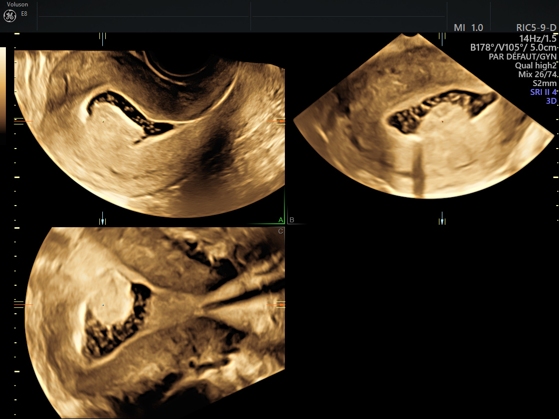
Submucosal fibroid enhanced with 3D Sonohysterography
One of Voluson’s biggest innovations is Radiantflow™. Radiantflow uses advanced algorithms to add height & depth information to color Doppler signals, to provide a 3D-like appearance with less flash and enhanced vessel boundaries. This can help improve your ability to visualize vascularization in uterine anatomy and pathology, for instance, to help distinguish polyps from fibroids. The advanced feature highlights the contours of the vessels in 3D-like appearance, providing valuable information about blood flow.
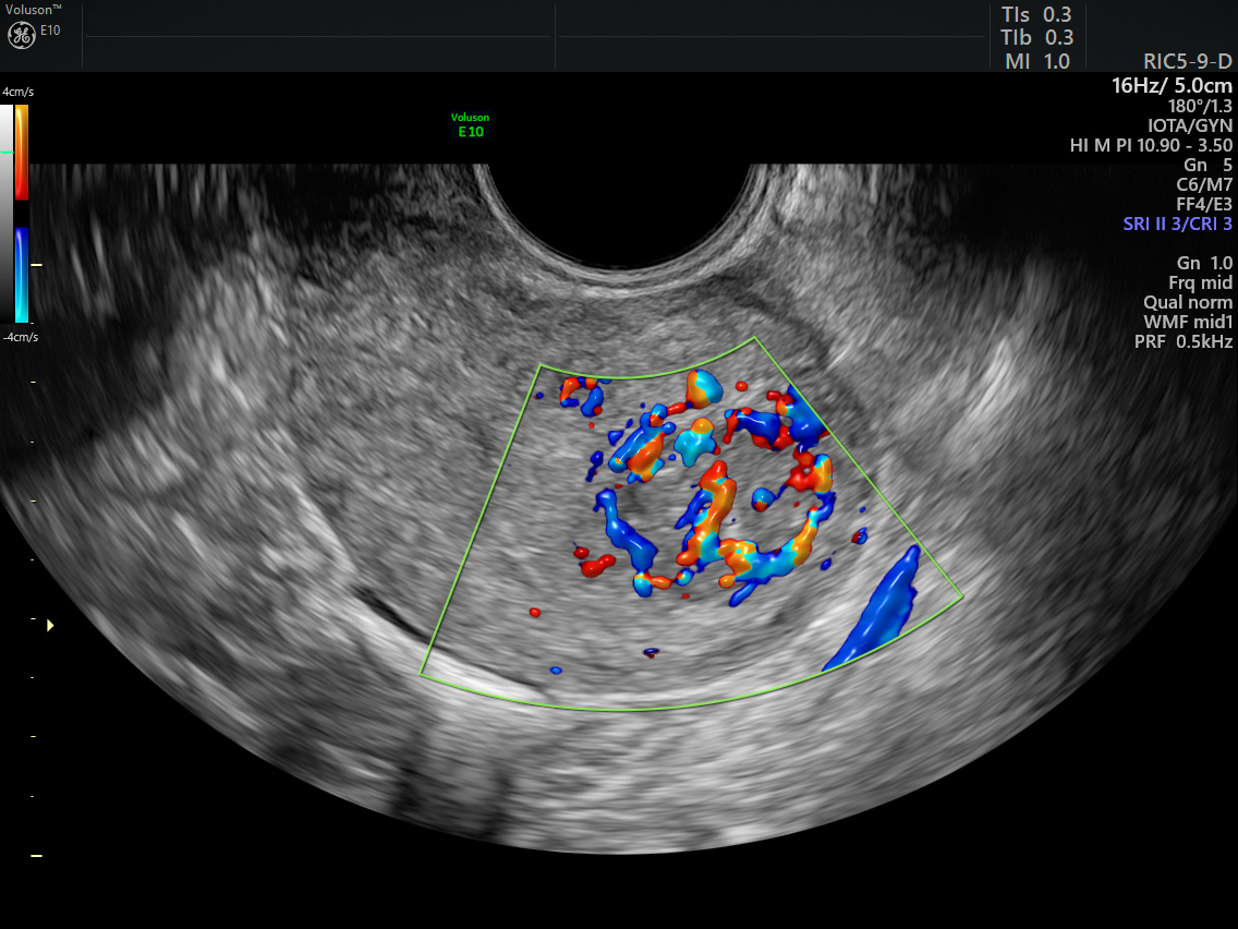
Uterine fibroid vasculature enhanced with Radiantflow
“Is it easier to see fibroids with Radiantflow? Absolutely. It hits you in that third dimension. Being able to assess blood flow can really contribute to your diagnostic capabilities. So, it’s ultrasound first—and with that information, maybe ultrasound only,” Dr. Platt attested.
The ability to effectively evaluate the blood supply of suspected fibroids is critical, especially in post-menopausal women. That’s because increased blood flow and irregular vascularization could be a sign of cancer.3 Monitoring any growth with ultrasound is also vital because fibroids often shrink after menopause when estrogen levels drop. While uterine sarcomas are rare—with only .5 to 3.3 cases per 100,000 women each year—these tumors are aggressive, with poor survival rates.5 It’s a serious risk for women that increases after age 50,6 and only reinforces the need for an early and accurate diagnosis.
Back in Amsterdam, Paulien knows the facts about fibroids as she inches closer towards menopause. And even though she’s found a few more fibroids over the years, Paulien stands by her doctor’s diagnosis. So, for now, it’s just a waiting game.
“For me, it’s important to keep monitoring with ultrasound as I go into menopause,” Paulien said. “I have high hopes they will start shrinking, but we will keep watching them.”
-
Learn how ultrasound can best be used in uterine fibroid treatment, diagnosis, classification and monitoring.
-
Learn how ultrasound-guided radiofrequency ablation, an alternative to surgery that uses heat to destroy tissue, can effectively treat fibroids.
-
What is ultrasound used for? Read our collection of FAQs.
-
A 3D ultrasound can help gynecologists identify potential causes of pelvic pain before recommending expensive surgical procedures or additional testing.
-
By catering to the concerns of women of fertility age, gynecologist can develop strong relationships that can last well past menopause.
-
Postmenopausal women have unique gynecological needs and concerns, many of which can be assessed with 3D ultrasound.
-
Gynecological care has come a long way, and 3D ultrasound empowers gynecologists to push the field into even more advanced territory.
Borah B., Nicholson W., Bradley L., Stewart E., The Impact of Uterine Leiomyomas: A National Survey of Affected Women, Am J Obstet Gynecol. 20132013 Oct; 209(4): 319.e1–319.e20.
Baird DD, Dunson DB, Hill MC, et al. High cumulative incidence of uterine leiomyoma in black and white women: ultrasound evidence. Am J Obstet Gynecol 2003; 188:100.
Buttram VC Jr, Reiter RC. Uterine leiomyomata: etiology, symptomatology, and management. Fertil Steril 1981; 36:433.
Serden SP, Brooks PG. Treatment of abnormal uterine bleeding with the gynecologic resectoscope. J Reprod Med 1991; 36:697.
Brooks SE, Zhan M, Cote T, Baquet CR. Surveillance, epidemiology, and end results analysis of 2677 cases of uterine sarcoma 1989-1999. Gynecol Oncol 2004; 93:204.
Hosh M, Antar S, Nazzal A, et al. Uterine Sarcoma: Analysis of 13,089 Cases Based on Surveillance, Epidemiology, and End Results Database. Int J Gynecol Cancer 2016; 26:1098.

