

Adnexal masses and ovarian lesions create a clinical dilemma. Without an established tissue diagnosis, providers must learn to rely on a combination of visual findings on ultrasound — power Doppler color scores, flow patterns and descriptive terms — to piece together a plan for either conservative management or surgical intervention.
An accurate and early diagnosis of ovarian cancer means treatment can significantly improve outcomes. But without effective biomarkers for mass screening, ovarian cancer is typically diagnosed in advanced stages. It has the highest mortality rate of the five gynecologic cancers, and is often referred to as "the silent killer."
Luckily, several regional and national scoring systems such as the International Ovarian Tumour Analysis (IOTA) group's Simple Rules are improving the early detection of ovarian cancer. All interface with transvaginal ultrasound, which captures tissue vascularity in real-time for accurate evaluation and is the preferred imaging tool because of its low cost and lack of radiation exposure.
The IOTA Group: Identifying Ovarian Malignancies
According to the Centers for Disease Control and Prevention, ovarian cancer accounts for only 3 percent of cancers in women in the United States; more large studies are required to establish the efficacy of protocols. The IOTA group, formed in 1999, is a collaboration between 50 centers around the world that combats this problem by developing common terminology and algorithms for the detection and care of ovarian malignancies and adnexal tumors.
By establishing a common, standardized terminology, IOTA helps clinicians everywhere better communicate findings, compare studies and collaborate. The preoperative risk classification term set known as the IOTA Simple Rules was developed through a combination of pattern recognition and color Doppler and performs better than previous scoring systems.
Under the IOTA classification system, findings are defined as:
- Unilocular, unilocular-solid, multilocular, multilocular-solid and solid tumors
- Anechoic, low level, ground glass, hemorrhagic or mixed cyst contents
- Solid material, solid papillary structures or wall irregularity (presence and size)
- Vascularity
- Shadows
- Ascites
3D ultrasound allows for superior evaluation of papillary structures, cyst walls and tumor volume. The 3D volume data sets can be obtained in a few seconds, stored and reviewed at a later time. These abilities mean this technology lends itself well to consultations and research, while reducing reliance on static, conventional two-dimensional planes that may not include all vital information.
The IOTA rules are a built-in feature of some ultrasound equipment. Training in this equipment is essential for learning how to use this classification system effectively.
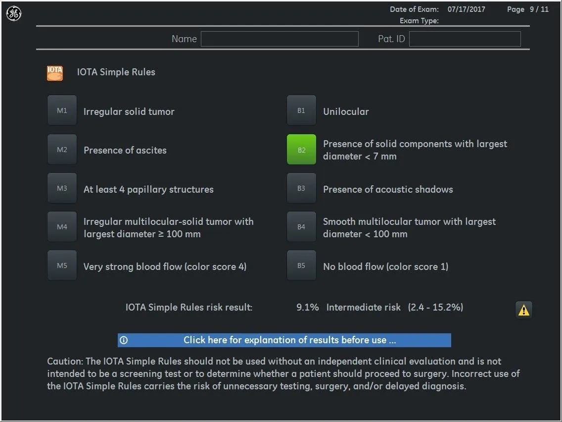
IOTA Simple Rules scan assistant feature on Voluson sytems
IOTA and Power Doppler
Angiogenesis is an important component of growth and metastasis of all malignancies, including ovarian malignancies. Recognizing the differences in the angiogenic nature of benign and malignant tumors is vital. Doppler can help identify markers like giant capillaries, arteriovenous shunts and tumor lakes that characterize new vessels in malignant lesions.
Power Doppler is three times more sensitive than conventional Doppler at detecting blood flow, making it ideal for visualizing complex vascular architecture dispersed throughout a lesion. This technology also displays the strength of the signal in color.
Due to the nature of neovascularization in malignancies, spectral Doppler may also be useful, as the vascular resistance index (RI) and pulsatility index (PI) is lower when compared to benign tumors. According to the IOTA models criteria, it is important to set the color pulse repetition frequency (PRF) at 0.3 kHz when measuring these indices.
The IOTA subjective color scoring system allows rapid assessment of the vascularity of a lesion:
- Color Score 1: No blood flow can be found in the lesion
- Color Score 2: Minimal flow can be detected
- Color Score 3: Moderate flow
- Color Score 4: Very strong blood flow
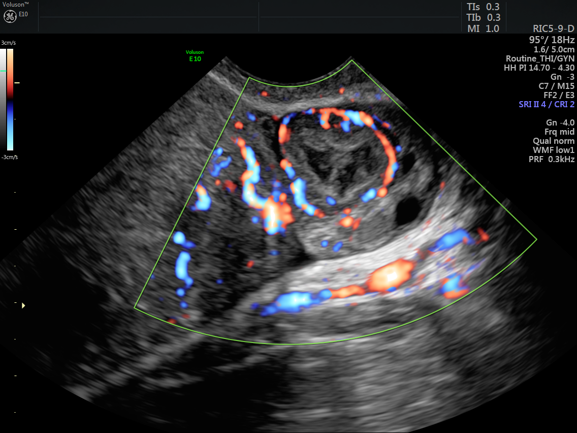
Ultrasound image of ovary demonstrating a color score of 4
Early diagnosis of ovarian malignancy is critical for improving patients' survival rates and quality of life. Using standardized terminology, and understanding the application of ultrasound and power Doppler findings in the differentiation and diagnosis of benign versus malignant ovarian tumors, can help clinicians plan patient care and improve the odds of diagnosing malignant ovarian lesions early on.

