Congenital Heart Defects: What if a Baby Has a Hole in Their Heart?
Congenital heart disease is the most common fetal birth defect, says an article in Indian Heart Journal, occurring in about 1 percent of all live, full-term pregnancies. Congenital heart defects include a range of structural abnormalities, but there are two main congenital septum defects that can occur:
Atrial septal defect (ASD) occurs when there is a persistent hole in the septum that divides the left and right atria (upper chambers) of the heart. This defect can vary in size and may close on its own.
Ventricular septal defect (VSD) is a hole in the muscular wall between the left and right ventricles (lower chambers) of the heart.
This article will examine these defects, their causes, symptoms, treatment options and more.
Causes and Diagnosis of Fetal Cardiac Septum Defects
The formation of the fetal cardiovascular system is a complex process involving both environmental and genetic components — and it is not completely understood why certain defects occur. Some genetic mutations are linked to defects of the heart; for example, 50 percent of babies with Down syndrome also have heart defects, according to the Centers for Disease Control and Prevention. However, risk factors for congenital heart defects can only be identified in 10 percent of those cases.
Certain maternal health conditions can increase the risk of having a baby with a congenital heart defect. These can include preexisting diabetes, rubella, lupus, maternal phenylketonuria and obesity. There are also some medications and lifestyle conditions that can lead to fetal heart defects, such as smoking or drinking alcohol during pregnancy.
Whether or not these risk factors are present, the fetal heart should always be examined carefully and closely on routine obstetric ultrasound exams. ASDs and VSDs are usually diagnosed with an ultrasound of the heart, either prenatally or postnatally.
Prenatal screening can be done with a maternal blood test to determine if any chromosomal abnormalities are present that could indicate an increased risk for cardiac defects. Some cardiac defects can be identified as early as 12 weeks of gestational age, but others cannot be identified until the second or third trimester, and some can only be confirmed after birth. Postnatal suspicion of a cardiac defect can occur if a doctor hears a heart murmur or if low blood oxygen is measured in the newborn. An echocardiogram will then be performed to determine what the defect could be.
Symptoms and Management of a Confirmed Fetal Heart Defect
When a fetal heart defect is identified or suspected, overall care will be transferred to a multidisciplinary center where specialized physicians are experienced in caring for obstetric patients and newborns with these conditions. A high-risk obstetrician will also become part of their care team. During the remainder of the pregnancy, the wellness of the fetus will be closely followed using weekly or biweekly obstetric ultrasounds and nonstress tests.
Prenatally, a fetus typically generally does not suffer from ill effects from a septal defect because the pressures in both the left and right sides of the heart are equal, resulting in blood flowing normally. After birth, the symptoms and problems that develop will depend on the size and location of the heart defect. The diagnosis will be confirmed with an echocardiogram for the newborn. Depending on the severity of the findings, the baby will either be discharged with their parents and scheduled for follow-up appointments or be admitted into the neonatal intensive care unit for treatment.
Confirming the symptoms and diagnosis of the heart defect is important to do postnatally because a newborn's cardiovascular system changes following birth, resulting in new blood flow patterns. One contributing factor is that, prenatally, the pressure in the fetal lungs is very high. But shortly after birth and during the first few months of life, the pressure in a baby's lungs decreases greatly.
When a VSD is present, this results in blood flowing in the direction of least pressure, so more blood flows into the lung vessels than is normal. For a time, a newborn's heart can compensate by pumping harder and faster, but this is an inefficient, short-term solution that will eventually lead to symptoms of increased respiration, difficulty feeding and limited weight gain or even weight loss.
When an ASD is present, the defect will result in abnormal left-to-right shunting of blood in the heart. If the septal defect is 10mm or smaller, the right heart experiences no enlargement or very minimal enlargement. But in the presence of larger septal defects, the right heart will be affected by a volume and pressure overload that causes enlargement. Longstanding shunting of blood through an ASD can lead to right heart hypertrophy, pulmonary hypertension and the development of pulmonary vascular disease.
Treatments and Surgical Options
Some VSDs will never be large enough to cause symptoms or even secondary effects in the heart. In these cases, treatment or surgery is unnecessary. Large VSDs, however, will start to negatively affect the heart. Medication can help to manage and lessen symptoms. These children will be followed by a cardiologist, who will closely monitor the heart defect and the course of treatment.
If medication does not work well enough or if the defect is very large and leads to uncontrollable symptoms, surgery may be considered. The VSD may be patched, closing the hole. This is typically done when the baby is 3–6 months old and may involve several days of recovery in the hospital. These surgical repairs have been shown to be very effective and should never need to be repeated. However, it is recommended that these children continue to regularly visit a cardiologist throughout their life to ensure their condition remains stable.
Long-term survival rates from a VSD are very high, and children with them are usually able to participate in typical activities — even sports. It is very rare for complications to develop later in childhood or adulthood.
Identifying Fetal Heart Defects with Ultrasound
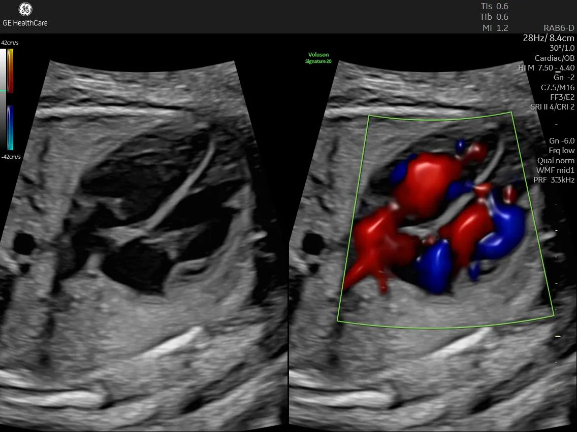
When evaluating the heart for potential defects, fetal ultrasound is used — mostly in the second and third trimesters, when the fetal heart is large enough to be clearly visualized. Standard planes of view are used to assess both the whole heart relative to its surroundings as well as structures within the heart, such as the septum. The classic four-chamber heart view is taken in the short-axis plane through the fetus's anterior abdominal wall. It is used to assess the overall heart's shape, size and position within the thorax as both the left and right atria and left and right ventricles can be seen together.
A lateral view (through either the left or right side of the fetal abdomen) is used to clearly identify and assess the septum. This angle must be perpendicular to the septum because it provides a better angle of the heart and prevents the drop-out effect that obscures most of the septum in the four-chamber view. The atrial septum is the upper third of the overall septum dividing the left and right atria and appears thinner and more membranous. The ventricular septum is the lower two-thirds of the overall septum, which divides the left and right ventricles and appears thicker and more muscular.
Typically, a 2D ultrasound assessment is completed first to evaluate the echogenic septum and identify gaps or defects. The image must be zoomed in so that the image of the heart fills as much of the screen as possible as well as optimized for clarity. Color and/or power doppler can also be used to determine if blood flow is moving through a defect in the septum.
Recent advances in 3D and 4D ultrasound technology have allowed for more detailed images and diagnoses of both smaller and more complex abnormalities of the heart and other organs.
Skill and Technology Lead to Better Fetal Outcomes
During obstetric ultrasound exams, it is critically important to thoroughly assess all parts of the fetal heart and septum for abnormalities. This often involves waiting for the fetus to be in the correct position and holding still long enough to allow for adequate interrogation.
Patience and experience as well as the use of the correct ultrasound technologies will ensure that even the smallest of heart defects can be identified — allowing for the best planning and treatment available for each newborn and their family.

